17 Disorders of the Vagina and Vulva
I. VULVAR DISCHARGE
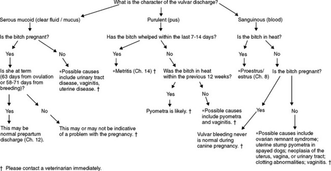
Discharge exuding from the vulva of bitches may arise from the uterus, cervix, vagina, or urinary tract (Figure 17-1). Localization of the source of the discharge often requires diagnostic testing of the reproductive tract and urinary tract at the same time. Urine is collected from the urinary bladder by cystocentesis, in which a needle is passed directly into the urinary bladder through the body wall. This prevents contamination of the urine with material from the vagina. Other diagnostic tests that allow localization of the source of vulvar discharge include examination of the vagina with a gloved finger or lighted scope, radiographs, ultrasound, submission of samples for culture, and examination of samples from the urinary or reproductive tracts microscopically to assess for the presence of blood, inflammatory cells, or abnormal cells as might be seen in animals with neoplasia. Finally, apparent vulvar discharge may arise from the skin around the vulva or may be expressed anal glands.
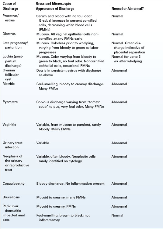
Vulvar discharge may be normal or abnormal. Determination of whether or not vulvar discharge is normal and localization of its source require physical examination and appropriate diagnostic testing (Table 17-1).
II. VAGINAL ANOMALIES
A. DEVELOPMENT
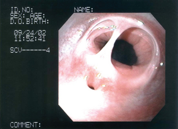
Vaginal septa are pillars or walls of tissue that form where dissolution of tissue normally would have occurred. A single pillar of tissue may be present just cranial to the urethral papilla (Figure 17-2) or the entire vaginal vault may be split. The split may be symmetrical or asymmetrical.
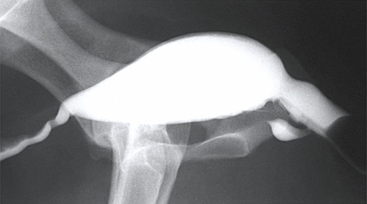
Vaginal strictures are 360-degree constrictions. These usually occur just cranial to the urethral papilla but also are described at the level of the vulvar lips and elsewhere in the vagina. There is a normal narrowing cranial to the urethral papilla, the cingulum, which should not be confused with a pathologic stricture (Figure 17-3).
III. VAGINAL PROLAPSE
A. DEVELOPMENT
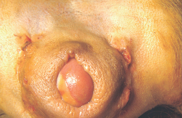
Vaginal prolapse is an overreaction of vaginal tissue to normal hormone production during proestrus and estrus. It is normal for vaginal tissue to become edematous and enlarged during proestrus because of the presence of elevated concentrations of estrogen in blood (see Chapter 9). In bitches that exhibit vaginal prolapse, the concentration of estrogen is not abnormal but the vaginal tissue becomes so enlarged that it falls out of the vaginal vault.
C. HISTORY AND CLINICAL SIGNS
Three stages of vaginal prolapse are described. Stage 1 is swelling of the perineal area (the area surrounding the anus and vulva). Stage 2 is protrusion of tissue from the floor of the vagina as a “tongue” of tissue (Figure 17-4). Stage 3 is protrusion of the entire circumference of the vagina as a “doughnut” of tissue. The urethral opening always is on the bottom of the prolapsed tissue and rarely is obstructed. Most bitches tolerate the prolapse with no other clinical signs.
< div class='tao-gold-member'>
Stay updated, free articles. Join our Telegram channel

Full access? Get Clinical Tree


