Chapter 28 DISORDERS OF THE PENIS AND PREPUCE
CONGENITAL DISORDERS
Persistent Penile Frenulum
Persistent penile frenulum is usually identified during the physical examination of a puppy that has been brought in for its first vaccinations (Fig. 28-1). Owners may also complain that the puppy urinates on its back feet or in other unexpected directions, which may cause secondary dermatitis of the rear leg from urine scald. Additional clinical signs may include inability to extrude the penis from the prepuce during penile engorgement, discomfort or pain with penile engorgement, and repeated licking of the preputial area. If the problem is not identified, the dog may associate pain with sexual excitement and secondarily develop reduced libido and unwillingness to mate.
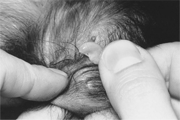
A diagnosis of persistent penile frenulum is made by identifying the abnormal band of tissue during visual examination of the penis and prepuce. The frenulum is usually avascular and can easily be transected with a scalpel blade after application of a topical anesthetic. Genetic implications for persistent penile frenulum are unknown, although Cocker Spaniels have been most commonly cited in the literature (Johnston et al, 2001).
Penile Hypoplasia
Penile hypoplasia is a rare disorder that has been reported in the Cocker Spaniel, Collie, Doberman Pinscher, and Great Dane (Johnston, 1989). Penile hypoplasia is a component of some intersex states (see page 893) and may be one of several reproductive abnormalities identified on physical examination. An abnormal karyotype has been identified in some animals with penile hypoplasia. A decrease in penile size may also occur in dogs castrated at an early age. Salmeri and colleagues (1991) found mixed breed dogs castrated at 7 weeks of age had immature genitalia, characterized by significantly smaller penile diameter, decreased size and radiodensity of the os penis, and immaturity of the prepuce, compared with male dogs castrated at 7 months of age or left intact. The mean penile diameter of the dogs castrated at 7 weeks of age was approximately half the penile diameter of the intact dogs; the penile diameter in dogs castrated at 7 months was intermediate between the other two groups.
Penile hypoplasia is usually asymptomatic and an incidental finding on physical examination. Urine pooling and infection inside the prepuce may develop if the dog also has a hypoplastic preputial opening (see page 954). The diagnosis of penile hypoplasia is based on physical examination. If it is identified, intersexuality should be suspected, and karyotyping (see page 893) may be indicated. No treatment is necessary in the asymptomatic dog. Enlargement of the preputial opening and surgical shortening of the prepuce may be necessary if urine pooling and infection are problems.
Hypospadias
Hypospadias is a congenital urogenital defect involving the external genitalia of males. It is characterized by an abnormal termination of the urethra ventral and posterior to the normal opening at the tip of the glans penis. Hypospadias is categorized as glandular, penile, scrotal, or perineal, depending on where the urethra opens (Hayes and Wilson, 1986). The perineal form is the most common and may represent a form of pseudohermaphroditism (see page 897). Hypospadias results from failure of fusion of the genital folds or genital swelling (or both) during fetal development, which may also cause abnormal development of the penis, prepuce, and/or scrotum. A short or deviated penis, malformed os penis, incomplete preputial closure, defects in the development of the scrotum, and other urogenital anomalies have been identified in dogs with hypospadias (see Fig. 24-14, page 898).
In addition to the visual abnormalities, clinical signs may include those related to urinary tract infection (e.g., dysuria, hematuria), urinary incontinence, and urine scalding. The diagnosis of hypospadias is made on visual inspection of the external genitalia and by catheterization of the urethra. The type of surgical correction required, if any, depends on the location and severity of the congenital defect. A thorough evaluation of the urogenital system should be completed before surgical correction is considered, because identification of additional abnormalities may alter the therapeutic plan. Penile amputation, ostectomy of the os penis, scrotal or perineal urethrostomy, or other reconstructive surgery may be necessary. Castration should always be performed because of the likely genetic implications of hypospadias, especially when the condition is present in conjunction with other developmental abnormalities (e.g., intersexuality, cryptorchidism). A familial occurrence in the Boston Terrier has been suggested (Hayes and Wilson, 1986).
Duplication of the Penis
Penile duplication (diphallia) is an extremely rare congenital anomaly that has been reported in a 5-month-old Pointer, a 6-month-old Poodle-cross, and a 5-month-old mixed breed dog with concurrent polymelia (Johnston, 1989; Zucker et al, 1993). All these dogs had multiple anomalies of the urogenital system (e.g., hydronephrosis, cryptorchidism, duplication of the urinary bladder and prostate gland). Diphallia may result from anomalous longitudinal duplication of the cloacal membrane, followed by ventral migration of primitive streak mesoderm around two cloacal membranes to form two genital tubercles. The diagnosis is made by visual inspection. There is no treatment.
ACQUIRED DISORDERS
Phimosis
The clinical signs depend on the severity of the defect. Complete congenital occlusion of the preputial orifice obstructs urinary outflow, resulting in neonatal death. Small openings may interfere with urination and cause accumulation of urine in the preputial cavity. Constant urine dribbling, an abnormal stream of urine during micturition, perpetual preputial swelling, and secondary bacterial infections and balanoposthitis may develop. More commonly, phimosis interferes with the extrusion of the penis during mating. Inability to copulate is quickly obvious. Pain may be associated with mating behavior, resulting in decreased libido.
Any predisposing factors should be corrected in dogs with acquired phimosis. The need for surgical intervention to widen the preputial orifice depends on the severity of the phimosis and the intended use of the dog. Therapy is probably not indicated in an asymptomatic dog not intended for breeding. Surgical reconstruction of the preputial orifice is indicated if the phimosis interferes with urination or with extrusion of the engorged penis in a dog intended for breeding. Congenital preputial stenosis has been observed in a litter of mixed breed dogs and in multiple related Golden Retriever litters, which suggests that this condition may be heritable (Johnston, 1989).
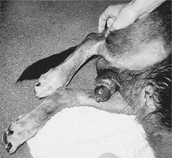
Paraphimosis
Paraphimosis is the presence of an engorged or edematous penis that cannot be retracted into the preputial sheath (Fig. 28-2). Interference with venous drainage of the cavernous tissues during engorgement results in progressive enlargement of the glans penis, interference with circulation to the penis, exposure desiccation, ischemic necrosis, and eventually the development of urethral obstruction and gangrene. Paraphimosis is most commonly associated with coitus, in which preputial hairs become entangled around the base of the glans penis, forming a restrictive band, or the protruding penis becomes trapped as the preputial orifice adheres to the penis and is pulled into the preputial cavity when the erection subsides. Other causes of paraphimosis include mild phimosis, foreign objects (e.g., rubber bands), fracture of the os penis, chronic priapism (i.e., penile engorgement without associated sexual excitement), trauma, neoplasia, and chronic balanoposthitis (Greiner and Zolton, 1980).
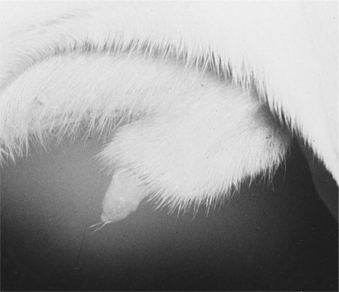
Other conditions may result in chronic exposure of the glans penis and may be mistaken for paraphimosis. These include paralysis of the retractor penis muscle, congenital deformation of the os penis, an abnormally large preputial opening, or congenital shortening of the prepuce (Fig. 28-3). These conditions can be differentiated from paraphimosis by the lack of an engorged penis and the length of time the problem is observed (i.e., chronic condition).
< div class='tao-gold-member'>
Stay updated, free articles. Join our Telegram channel

Full access? Get Clinical Tree


