27 Disorders of the Penis and Prepuce
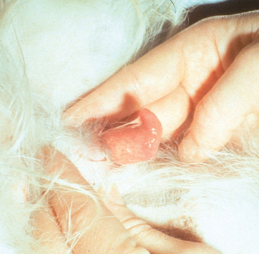
I. PERSISTENT PENILE FRENULUM
A. DEVELOPMENT
The penis and prepuce are joined during embryonic development by a tissue called the balanopreputial fold. This fold should dissolve, under the influence of testosterone, before birth. In some dogs, persistence of a portion of this fold leads to a permanent connection between the ventral portion of the penis and the prepuce, such that the penis cannot be completely extruded. The penile frenulum also may be associated with the penis only, usually causing ventral deviation of the tip of the penis as the frenulum attaches the tip to the shaft (Figure 27-1).
II. PHIMOSIS, PARAPHIMOSIS, AND PRIAPISM
A. DEVELOPMENT
Phimosis is the inability of the penis to be extruded from the prepuce (Figure 27-2). Paraphimosis is the inability of the nonerect, extruded penis to be withdrawn into the prepuce. Priapism is persistent erection and an inability of the erect, extruded penis to be withdrawn into the prepuce.
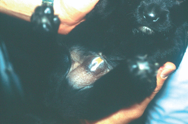
Figure 27-2 Phimosis.
(From Johnston SD, Root Kustritz MV, Olson PN: Canine and feline theriogenology, Philadelphia, 2001, Saunders.)
Phimosis can occur whenever there is narrowing of the external portion of the prepuce. It is uncommon and usually occurs as a congenital defect in puppies. Paraphimosis may occur because of a congenital defect in which the muscles allowing the penis to be drawn into the prepuce are dysfunctional or the external orifice of the prepuce is too small, but more often it occurs in older dogs that have historically been able to replace their penis within the prepuce. It may occur secondary to inflammation or injury of the penis. Most often, paraphimosis in older dogs has no identifiable cause. Priapism occurs with obstruction of normal outflow of blood from the engorged penis. This may occur secondary to neoplasia, thromboembolism (blood clot), neurologic defect, or unfortunate placement of sutures during castration.
C. HISTORY AND CLINICAL SIGNS
Dogs with phimosis usually have an inability to urinate or dribbling of urine. The puppy pictured in Figure 27-2 was unable to urinate unless the prepuce was compressed; urine would then spray out of a pinpoint opening present on the prepuce.
E. TREATMENT
Paraphimosis can be a frustrating disorder to treat. Although castration might be of benefit to some dogs, this disorder often occurs in dogs that already have been castrated. An underlying cause often cannot be identified. Treatment is aimed at maintaining the penile tissue in good condition and preventing the dog from licking at it excessively. The dog should be removed from situations that might cause excitement. If the dog is disciplined excessively for licking at the penis, it may regard that as a positive stimulus or reward; the owner is cautioned not to make corrections the dog finds attractive or attention getting. The penis should be cleaned and lubricated. If the penis has been damaged, it should be examined and, if necessary, repaired by a veterinarian before it is replaced. Some dogs respond well to medical therapy with oral progesterone-type drugs.
Stay updated, free articles. Join our Telegram channel

Full access? Get Clinical Tree


