Diseases of the Reproductive System

Chapter 10 Diseases of the Reproductive System


The female reproductive system consists of two ovaries and the female duct system, including the oviducts, uterus, cervix, vagina, and vulva (Fig. 10-1). The primary functions of this system are to provide eggs for fertilization and to protect the developing embryo during pregnancy. All of these structures are composed of tissue that is sensitive to hormones produced by the female.
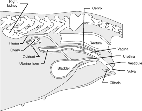


Hormones such as estrogen and progesterone act on the reproductive system to prepare it for pregnancy and to maintain that pregnancy. When the response to these hormones is abnormal, disease can result. Although not technically a part of the female reproductive system, the mammary glands are also reactive to hormonal abnormalities, and diseases involving them are frequently seen in small-animal practice.


The male reproductive system consists of two testicles and the male duct system, including the urethra, prostate gland, and penis (Fig. 10-2). Other structures often involved with disease processes are the scrotum and the prepuce. The main hormonal influence in the male system is testosterone, although abnormal estrogen levels can also affect the male reproductive system.
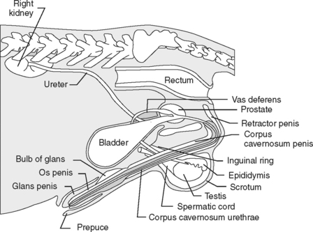


Diseases that involve the reproductive system are frequently seen in veterinary practice. These include vaginal disorders, uterine disorders, pregnancy disorders, lactation disorders, disease of the prostate, and neoplasia of the genital system and mammary glands.


FEMALE REPRODUCTIVE DISEASE


Pyometra


Pyometra is also frequently seen in small-animal medicine. Increasing levels of progesterone after ovulation result in hyperplasia and hypertrophy of the endometrial glands of the uterus. Inappropriate response results in cystic endometrial hyperplasia with accumulation of fluid within the uterine lumen. Progesterone also produces a decrease in myometrial contractions and predisposes the uterus to secondary bacterial infection (pyometra). The most common microorganism isolated in pyometra is Escherichia coli, although Staphylococcus spp., Streptococcus spp., Klebsiella, Pasteurella, Proteus, and Moraxella have also been implicated.


The development of pyometra appears to be the final stage of a continuum beginning with endometrial hyperplasia and progressing through cystic endometrial hyperplasia and endometritis. Animals presented for treatment tend to be middle-aged or older, within 60 days of their last estrous cycle.


Pregnancy Disorders


Disorders of pregnancy include fetal death and abortion/reabsorption, dystocia, inappropriate maternal behavior, mastitis, and puerperal tetany. Although other problems are associated with pregnancy and parturition, these are the most commonly seen problems in small-animal medicine. The normal gestation period for the dog and cat is between 62 and 65 days. Fetuses can be palpated about 25 to 36 days after breeding in the dog, and 21 to 28 days in the cat. Fetal skeletal mineralization can be detected radiographically at 45 days gestation. Ultrasonography can provide information on the status of the fetuses after about 20 days. It is difficult to determine the number of fetuses, especially in large litters.


Fetal deaths early in gestation result in reabsorption with no expulsion of uterine contents. Owners may report that the animal has “failed to conceive” after what they consider a successful breeding. Organisms such as Brucella canis, canine herpesvirus, feline infectious peritonitis, feline leukemia virus, and panleukopenia can produce fetal death or abortion.


Dystocia can be defined as difficulty in delivery of fetuses through the birth canal. The causes of dystocia can be divided into fetal factors, maternal factors, and combinations of both. Fetal factors include large fetuses (large puppy or kitten, fetal anasarca, or hydrocephalus) and abnormal positioning (transverse presentation). Breech presentation is not an abnormality in the bitch or queen. Maternal factors include a narrowed birth canal (developmental or trauma related) and uterine inertia (lack of coordinated contractions or exhaustion of the uterine musculature from prolonged contractions).


Dystocia


Aug 31, 2016 | Posted by in GENERAL | Comments Off on Diseases of the Reproductive System

Full access? Get Clinical Tree

Get Clinical Tree app for offline access