Diseases Formerly Known as Rickettsial: The Rickettsioses, Ehrlichioses, Anaplasmoses, and Neorickettsial and Coxiella Infections

CHAPTER 18 Diseases Formerly Known as Rickettsial


The Rickettsioses, Ehrlichioses, Anaplasmoses, and Neorickettsial and Coxiella Infections


Clinically Relevant Nomenclature


In the strictest sense, “rickettsial” infection refers to infection with organisms belonging to the genus Rickettsia. Historically rickettsial has been used to describe infection with organisms belonging to family Rickettsiaceae. The organisms in this family included obligately intracellular organisms with phenotypic characteristics of both viruses and bacteria. Several genera of bacteria belonged to the family, including (but not limited to) Rickettsia, Ehrlichia, Neorickettsia, and Coxiella. Recent genetic analyses of 16S rRNA genes, heat shock and surface protein genes, along with consideration of various phenotypic characteristics, have resulted in dramatic changes in the classification and ultimately the composition of the family Rickettsiaceae. Members of the genera Ehrlichia and Neorickettsia were entirely removed and placed into the family Anaplasmataceae. Like Ehrlichia and Neorickettsia, Coxiella has also been removed from the family Rickettsiaceae and has been placed in the more distant family Coxiellaceae.


As a result of these taxonomic rearrangements, the family Rickettsiaceae currently includes only the genera Rickettsia and Orientia, which are intracellular bacteria that live free in the cytoplasm or nucleus of vertebrate host cells. The family Anaplasmataceae includes intracellular organisms that reside within vacuoles of hematopoietic cells (erythrocytes, monocytes, neutrophils, and platelets) and includes (but is not limited to) the genera Ehrlichia, Anaplasma, and Neorickettsia.


Because these organisms no longer share the family name Rickettsiaceae, we propose to limit the term rickettsioses or rickettsial disease to disease caused by organisms in the genus Rickettsia. Accordingly in this manuscript, diseases caused by Ehrlichia, Anaplasma, Neorickettsia, and Coxiella are referred to as ehrlichioses, anaplasmoses, and neorickettsial and Coxiella infections, respectively.


Tickborne Rickettsioses, Ehrlichioses, and Anaplasmoses


Several species within the genera Rickettsia, Ehrlichia, and Anaplasma are transmitted to vertebrate hosts through tick vectors. Accordingly the geographic distribution of disease usually follows the distribution of ticks infected with and capable of transmitting a particular organism. It is clinically useful to consider the known geographic distribution of vectors and the disease-causing agents they carry when determining differential diagnoses for infection. For example, for a dog presenting with neutrophilic polyarthritis in Wisconsin, infection with Anaplasma phagocytophilum (transmitted by Ixodes scapularis) would be a more likely diagnostic consideration, whereas in the southern United States, Ehrlichia ewingii (transmitted by Amblyomma americanum) would be a more likely differential diagnosis. Therefore practitioners should ensure that tests used to diagnose tickborne diseases (e.g., the “tick panel”) are appropriate for organisms prevalent in their area of practice or relevant to a particular patient. The distribution of some tick vectors of clinical significance in the United States can be found at http://www.cdc.gov/Ncidod/dvrd/rmsf/Natural_Hx.htm and http://www.cdc.gov/mmwr/preview/mmwrhtml/rr5504a1.htm.


Despite these considerations, it is important to be aware that these organisms have been very difficult to study in the past because of their complex and highly specialized life cycles. Therefore information regarding their epidemiology and pathophysiology remains incomplete. Recent advancements in molecular biology have led to increased understanding of the spectrum of diseases caused by these agents, the extent of their geographic distribution, and the range of species that are capable of causing disease in people. It is important that veterinarians consider the possibility of infection with a novel vector-borne agent when pets present with atypical clinical signs.


Atypical clinical signs may also be caused by coinfection with multiple organisms. Coinfection with Ehrlichia, Bartonella, Rickettsia, and Babesia, or Anaplasma, and Borrelia has been documented and is likely attributable to coinfection in tick vectors transmitting the agents in a particular geographic location. Atypical clinical signs or failure to respond to appropriate therapy should prompt consideration for testing for additional agents.


Importantly, treatment of infection must begin before clinical confirmation of the causative infectious agent in most cases. Delayed or inappropriate antibiotic therapy results in increased morbidity and mortality. Doxycycline is the treatment of choice for adult animals. Fluoroquinolones are not recommended for treating ehrlichioses or anaplasmoses. Special considerations for the treatment of the pediatric patient are necessary.


Finally, it is important for the veterinarian to be aware that companion animals can serve as sentinels for some of these vector-borne diseases in people. Communication with owners and their physicians regarding a documented infection in a companion animal and the possibility of simultaneous or subsequent human exposure to the same infected vectors can be lifesaving. The role of companion animals as sentinels for disease is particularly important for Rickettsia rickettsii and Coxiella burnetii because these agents can cause serious morbidity or mortality in people. These organisms are also considered by the U.S. government to be potential weapons for use in bioterrorist attacks.


Rickettsioses: The Spotted Fever Group Rickettsia


The genus Rickettsia is divided into the spotted fever group (SFG) and the typhus group. The SFG Rickettsia includes important causes of emerging and reemerging infectious disease worldwide. These organisms are transmitted by arthropod vectors and primarily infect endothelial cells in vertebrate hosts. Many species of SFG Rickettsia have been associated with disease in people, with several being recognized very recently.


R. rickettsii and Rickettsia conorii cause Rocky Mountain spotted fever (RMSF) and Mediterranean spotted fever (MSF), respectively, in people. Because of their pathogenicity, these are the best characterized of the SFG Rickettsia. R. rickettsii also causes RMSF in dogs. Recently we documented infection with R. conorii in Italian dogs presenting with signs consistent with a spotted fever–like illness. Therefore R. conorii infection should be considered in dogs with a spotted fever–type illness in endemic areas. Evidence suggests that Rickettsia akari, Rickettsia japonica, and Rickettsia australis may also infect dogs. The role of these and other SFG Rickettsia as disease-causing agents in dogs is beginning to be elucidated.


As the most pathogenic and well-characterized SFG Rickettsia infecting dogs, R. rickettsii will be considered in detail here. Although cats can be seroreactive to R. rickettsii, clinical disease has not been well characterized. The ability of typhus group Rickettsia to cause disease in dogs and cats has also not been well characterized and will not be described here.


Rickettsia rickettsii and RMSF in Dogs


RMSF is a reportable disease in people in the United States. Reporting is also mandatory for dogs in some regions of the country. Natural and experimental infection in dogs has been well documented. As is the case in people, Dermacentor ticks are believed to be the primary vector for dogs in the United States. However, Rhipicephalus sanguineus, a one-host tick that prefers to feed on dogs, may also play a role in transmitting R. rickettsii to dogs. Dogs living outdoors are at increased risk for infection, and most canine cases of RMSF coincide with months of increased tick feeding activity. In dogs, a sex predisposition has not been substantiated across studies. Similarly, studies investigating whether infection is more likely in young dogs (aged less than 2 years) are conflicting. Whether purebred dogs may be at increased risk for infection is also controversial. Springer Spaniels with phosphofructokinase deficiency and German Shepherd Dogs may suffer from more severe disease. The majority of dogs with RMSF do not have a history of a tick bite.


RMSF is an acute illness, with the onset of clinical signs occurring within 2 to 14 days after transmission. In dogs, the most common signs are nonspecific and include fever, anorexia, and lethargy. Although common, it is important to note that fever is not always present. R. rickettsii infect endothelial cells and therefore induce vasculitis. The pathologic consequences of vasculitis include disorders of primary hemostasis and edema. Many of the clinical signs of RMSF reflect the presence of vasculitis and systemic inflammation involving several body systems. Ocular signs are common and may include discharge, scleral and conjunctival injection and hemorrhage, conjunctivitis, uveitis, retinal hemorrhage, and retinitis. Respiratory signs can include nasal discharge, epistaxis, tachypnea, dyspnea, and coughing. Gastrointestinal signs may include vomiting, diarrhea, and melena. Cutaneous and mucocutaneous abnormalities can include petechiae, ecchymoses, edema, hyperemia, and necrosis. Orchitis, scrotal edema, hyperemia, hemorrhage, and epididymal pain are often present in intact male dogs. Hematuria may also occur. Myalgia and arthralgia signal muscle and joint involvement. Central nervous system (CNS) signs can be focal or generalized and can include hyperesthesia, ataxia, vestibular signs, stupor, seizures, and coma. Dramatic weight loss, disproportionate to the amount of time the dog has been ill, has also been described. Severe consequences of vasculitis and increased vascular permeability include edema, microvascular hemorrhage or thrombosis, hypotension, and gangrene. As a result, oliguric renal failure, cardiovascular collapse, and brain death can occur terminally.


Typical hematologic findings in dogs include normocytic normochromic anemia and thrombocytopenia. Thrombocytopenia has been identified as one of the most consistent hematologic abnormalities. Thrombocytopenia likely occurs as a result of immune-mediated destruction and sequestration of platelets secondary to vasculitis. Although generally mild, thrombocytopenia can be severe. Leukopenia can occur in the first 48 hours of infection, after which leukocytosis, neutrophilia, monocytosis, lymphopenia, and eosinopenia can occur. Toxic change within neutrophils and metamyelocytes may be observed. Serum chemistry abnormalities can include hypoalbuminemia, azotemia, hyponatremia, hypocalcemia, increased alanine aminotransferase and alkaline phosphatase, mild hyperbilirubinemia, and bilirubinuria. Cerebrospinal fluid analysis may reveal a mild increase in protein and cells with a neutrophilic pleocytosis occurring acutely, followed by a mononuclear pleocytosis later in the course of the disease. Proteinuria may be present secondary to glomerular or tubular damage. Joint fluid analysis is consistent with neutrophilic polyarthritis. Animals rarely develop disseminated intravascular coagulation; however, abnormalities in fibrinogen levels (increases or decreases), elevated fibrin/fibrinogen degradation products, decreased antithrombin, and prolongation of activated partial thromboplastin time or activated clotting time can occur. Thoracic radiographs may show an increased interstitial pattern reflecting pneumonitis.


Because of the nonspecific nature of the clinical abnormalities associated with RMSF, confirmatory testing is necessary for diagnosis and requires serologic testing, direct immunofluorescent testing for R. rickettsii antigen in biopsy specimens, and/or polymerase chain reaction (PCR) testing for rickettsial DNA.


Serology is commonly used to diagnose SFG rickettsiosis. Importantly, titers can be negative early in infection, and antibodies can be detected in animals with no history of illness in endemic areas. Therefore seroconversion (a fourfold increase or decrease in antibody titer) must be documented to confirm acute infection. An acute phase sample should be obtained at presentation and a convalescent sample obtained 2 to 3 weeks thereafter. Antibody titers greater than 1 : 1024 obtained 1 week or more after the onset of clinical signs have also been used to diagnose infection. However, it is important to note that high antibody titers can persist in dogs long after recovery, and therefore high titers in the absence of seroconversion must be interpreted in the context of the individual patient. Notably, serologic cross-reactivity among SFG Rickettsia occurs; therefore the species of infecting SFG Rickettsia is presumed to be R. rickettsii.


Immunohistochemistry and Gimenez staining of tissue samples can also be used to demonstrate SFG Rickettsia and therefore can confirm the diagnosis early in the disease course. Importantly, neither determines the species of infecting SFG Rickettsia.


Demonstration of rickettsial DNA using PCR also confirms infection. R. rickettsii infect endothelial cells and circulate in the bloodstream in very low numbers. Therefore detecting the organism in blood can be challenging. Recently a very sensitive PCR was specifically developed to test dog blood for the presence of SFG Rickettsia DNA (Vector Borne Disease Diagnostic Laboratory, North Carolina State University). Importantly, this test can detect Rickettsia in blood before seroconversion and may be useful in confirming infection early. However, it is important to note that rickettsial DNA cannot be demonstrated in the blood of all acutely infected dogs, and a negative test does not indicate a lack of infection. Notably, the species of infecting Rickettsia can be confirmed by sequencing the gene product using this test.


Treatment must begin before results of confirmatory testing are available and is based on a high index of clinical suspicion by the veterinarian. Response to appropriate antibiotic therapy is rapid and dramatic. The consequences of delayed or missed diagnosis and inappropriate antibiotic therapy can be severe morbidity or death. Doxycycline (5 mg/kg twice a day for 7 to 14 days) is the treatment of choice for RMSF. Chloramphenicol is also effective, but studies in people suggest it may be less effective than doxycycline. The undesirable side effect of bone marrow suppression precludes its use in people and should be considered as a less likely but potential side effect in dogs. Owners should wear gloves and avoid inhaling the drug if it is used to treat RMSF in dogs. Enrofloxacin is also effective in treating RMSF in dogs. Notably, studies suggest chloramphenicol may not be as effective as doxycycline in inhibiting the growth of A. phagocytophilum, Ehrlichia canis, or Ehrlichia chaffeensis in vitro. In addition, fluoroquinolones are less effective in inhibiting the growth of E. canis or E. chaffeensis in vitro and do not clear infection in dogs infected with E. canis. Furthermore, chloramphenicol is inferior to doxycycline for treating human monocytic ehrlichiosis (HME) caused by E. chaffeensis. Diseases caused by these organisms in dogs are often differential diagnoses for unconfirmed cases of RMSF presenting acutely. In addition, they may act as coinfecting agents. Therefore doxycycline offers several advantages over other antibiotics that have activity against R. rickettsii. Treatment considerations in the pediatric patient are covered later in this chapter.


Other treatment for RMSF consists of supportive care, which may include cautious use of colloids and fluid therapy (exacerbation of edema with fluid therapy is a concern). Treatment of coagulopathies may be necessary, but clinical and hematological improvement following initiation of doxycycline is generally rapid. Short-term immunosuppression with glucocorticoids may be considered in dogs with severe thrombocytopenia with a suspected immune-mediated component. Most clinical signs resolve rapidly after instituting appropriate antibiotic therapy, often within 12 to 48 hours. Residual neurologic signs or gangrenous lesions requiring surgery may occur in severe cases. If rapid response to treatment is not observed, coinfection with Babesia, Ehrlichia, Bartonella, or other organisms or other alternative diagnoses should be considered.


Immunity to natural infection with R. rickettsii appears to be permanent. Minimizing tick exposure, routine use of acaricides, and removing ticks from dogs daily are effective means of prevention.


Dogs are sentinels for the disease in people. Therefore veterinarians should notify owners and their physicians when RMSF is diagnosed in a dog (Box 18-1). Owners should be instructed with regard to proper tick removal, and crushing the tick should be avoided to prevent inadvertent exposure to infected hemolymph. Hands should be washed immediately after removal. Contact with infected dog blood by veterinarians and support staff could result in transmission of the organism, although this risk is presumably small. Wearing gloves, washing hands, avoiding aerosolization, and using zoonotic warning labels are recommended for handling samples from suspect cases.


Ehrlichiosis and Anaplasmoses


Members of the genera Ehrlichia and Anaplasma are obligate intracellular parasites that reside in vacuoles within hematopoietic cells in clusters (morulae). The organisms are transmitted to mammals through the salivary glands of ticks while obtaining a blood meal. Recently there has been a large shift in the taxonomic organization of these organisms that is relevant to the small animal practitioner. Genetic analysis has shown that the agents formerly known as Ehrlichia equi and the agent of human granulocytic ehrlichiosis (HGE) are variants of the species A. (formerly Ehrlichia) phagocytophilum; thus they have been renamed accordingly. Furthermore, Ehrlichia platys has been reclassified and has been renamed Anaplasma platys. Finally, Ehrlichia risticii is now classified as Neorickettsia risticii.


Although the disease syndrome caused by these organisms is similar, there are apparent differences in their epidemiology, pathophysiology, and response to treatment. For example, the agents that cause granulocytic anaplasmosis and ehrlichiosis (A. phagocytophilum and E. ewingii) infect neutrophils and appear more likely to cause polyarthritis than the agents of monocytic ehrlichiosis. Furthermore, infections with A. phagocytophilum usually appear to induce more acute disease that responds more rapidly to doxycycline therapy than monocytic ehrlichiosis caused by E. canis or E. chaffeensis. Notably, E. ewingii causes chronic infection in dogs, whereas the extent to which A. phagocytophilum induces chronic infection is unknown. It is helpful to keep these differences in mind when determining appropriate diagnostic and therapeutic plans for affected patients.


The following is a brief review of Ehrlichia and Anaplasma species known to cause disease in dogs and cats.


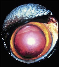
Canine Monocytic Ehrlichiosis Caused by Ehrlichia canis or Ehrlichia chaffeensis


Ehrlichia canis


E. canis is a gram-negative obligately intracellular organism that resides in vacuoles within mononuclear cells. It is transmitted by R. sanguineus, the brown dog tick. R. sanguineus feeds on canid hosts during all three stages of its life cycle. This tick requires dry environments to maintain water balance. Therefore it often infests isolated dry environments of homes and kennels. Although R. sanguineus prefers to feed on canids, its proximity to humans results in occasional feeding, and therefore disease transmission to this less-desirable host is possible.


R. sanguineus and E. canis have a worldwide distribution. In general, infection appears to be more common in warmer locales. Seroprevalence studies sampling dogs from multiple regions of the United States suggest the disease occurs in most states but is more common in the Southeast. Most cases of HME are caused by infection with E. chaffeensis. Because serologic cross-reactivity between E. chaffeensis and E canis occurs, previous reports of HME attributed to E. canis have been disputed. However, recently infection with E. canis has been confirmed in people with HME using molecular techniques.


The manifestations of disease caused by E. canis in dogs can be characterized as acute, subclinical, and chronic, although distinguishing these phases in the clinical setting may be difficult. It has been hypothesized that variability in the manifestation of clinical disease is likely caused by E. canis strain differences, the presence of coinfecting agents, the immune status of the patient, and host genetic factors. Increased susceptibility and disease severity have been reported in German Shepherd Dogs.


After transmission, an incubation period of 8 to 20 days occurs before clinical signs develop. The acute phase of illness lasts 2 to 4 weeks. Clinical signs include anorexia, lethargy, fever, lymphadenomegaly, splenomegaly, hepatomegaly, weight loss, scleral injection, and oculonasal discharge. Neurologic abnormalities may also occur. The most consistent and prominent hematologic abnormality is thrombocytopenia. Evidence suggests the thrombocytopenia associated with E. canis infection is caused by vasculitis, immune-mediated destruction, and sequestration. Other hematologic abnormalities that occur during the acute phase include leukopenia and nonregenerative anemia. Transient hypoalbuminemia and proteinuria have been documented during the acute phase in experimentally infected dogs. Glomerular pathology, including minimal change disease and glomerulonephritis, has been associated with proteinuria during this phase. Dogs may recover from the acute phase and clear the infection or progress to the subclinical phase.


The subclinical phase lasts months to years, and dogs appear healthy during this time. Thrombocytopenia can persist in the subclinical phase. Neutropenia and lymphocytosis also occur. Hyperglobulinemia appears to be common during the subclinical phase of infection. Although usually polyclonal in nature, the gammopathy associated with E. canis infection can also be monoclonal.


During the chronic phase, nonspecific signs such as weight loss, lethargy, and anorexia can recur. Disorders of primary hemostasis caused by thrombocytopenia and platelet dysfunction are prominent and include epistaxis, petechial or ecchymotic hemorrhages, hematuria, and melena. Fever, lymphadenopathy, and splenomegaly may also be present. Dyspnea resulting from pneumonitis can also occur. Uveitis is common in dogs with E. canis infection. Other ocular abnormalities include corneal ulceration, hyphema, retinal hemorrhage, necrotic scleritis, decreased tear production, and orbital cellulitis (Figure 18-1). Importantly, ocular abnormalities can be the only presenting clinical sign. Neurologic signs include ataxia, seizures, head tilt, nystagmus, and conscious proprioceptive deficits. Hematologic findings include nonregenerative or rarely Coombs-positive immune-mediated hemolytic anemia. Like the acute and subacute phases of E. canis infection, thrombocytopenia can persist in the chronic phase. White blood cell counts can be normal, increased, or decreased, and pancytopenia may be present. Large granular lymphocytosis, which can be confused with leukemia, is noted on blood smears in some cases. Examination of bone marrow may reveal hypoplasia of the erythroid, myeloid, and/or megakaryocytic cell lines. Hyperplasia, particularly of the megakaryocytic cell line, may occur during the acute phase of the disease. Interestingly, bone marrow examination can sometimes reveal plasma cell hyperplasia, which can be confused with neoplasia.


Stay updated, free articles. Join our Telegram channel

Sep 11, 2016 | Posted by in SMALL ANIMAL | Comments Off on Diseases Formerly Known as Rickettsial: The Rickettsioses, Ehrlichioses, Anaplasmoses, and Neorickettsial and Coxiella Infections

Full access? Get Clinical Tree

Get Clinical Tree app for offline access