Chapter 9 Digestive system
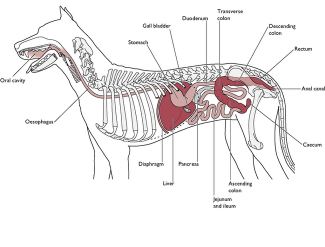
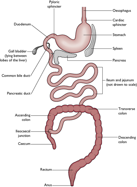
The digestive system (Figs 9.1, 9.2) consists of the following parts:
There are also several accessory glands, without which the digestive process cannot be completed:
The oral cavity
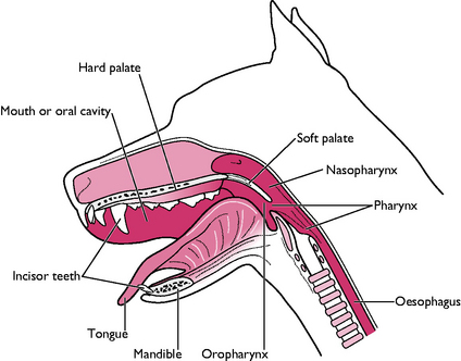
The oral cavity (Fig. 9.3) is also known as the mouth or buccal cavity and contains the tongue, teeth and salivary glands. The function of the oral cavity is as follows:
The oral cavity is formed by the following bones of the skull:
The upper and lower jaws are linked by skin, forming the cheeks, under which lie the muscles of mastication (see Ch. 4). These muscles lie over the temporomandibular joint and give strength to the biting action. The entrance to the mouth is closed by the lips, composed of muscle covered in skin. The upper lip is split vertically by a division known as the philtrum (see Ch. 8, Fig. 8.1).
The entire oral cavity is lined by a layer of mucous membrane. It is reflected on to the jawbones, forming the gums. The mucous membrane covers the hard palate and extends over a flap of soft tissue at the back of the oral cavity – this is the soft palate, which extends caudally between the oral and nasal cavities and divides the pharynx into the oropharynx and nasopharynx (Fig. 9.3) (see also Ch. 8).
The tongue
The functions of the tongue are:
The tongue lies on the floor of the oral cavity and is made of striated muscle fibres running in all directions. This enables the tongue to make delicate movements. The muscles are attached at the root of the tongue to the hyoid bone (see Ch. 8) and to the sides of the mandibles. The tip of the tongue is unattached and very mobile.
The tongue is covered in mucous membrane. The dorsal surface is thicker and arranged in rough papillae, which assist in control of the food bolus and in grooming. Some papillae are adapted to form taste buds, mainly found at the back of the tongue. Taste buds are well supplied with nerve fibres, which carry information about taste to the forebrain (see Ch. 5). Running along the underside of the tongue are the paired sublingual veins and arteries.
The teeth
Structure
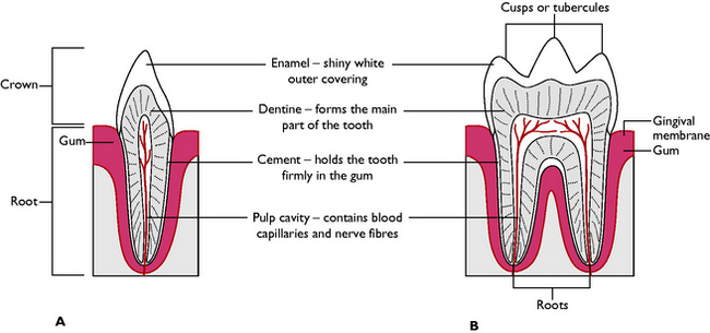
All teeth have a basic structure (Fig. 9.4). In the centre of each tooth is a pulp cavity. This contains blood capillaries and nerves, which supply the growing tooth. In young animals the cavity is relatively large but, once the tooth is fully developed, it shrivels and contains only a small blood and nerve supply. After a tooth has stopped growing the only changes occurring will be due to wear.
Function
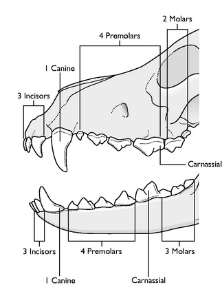
The teeth of a carnivore are adapted to shearing and tearing the flesh off the bones of their prey. There are four types of tooth, which are classified by their shape and position in the jaw (Fig. 9.5). This is summarised in Table 9.1.
Table 9.1 Tooth types and functions
| Type | Position and shape | Function |
|---|---|---|
| Incisor (I) | Lie in the incisive bone of the upper jaw and in the mandible of the lower jaw; small, pointed with a single root | Fine nibbling and cutting meat; often used for delicate grooming |
| Canines (C) – ‘eye teeth’ | One on each corner of the upper and lower jaws; pointed, with a simple curved shape; single root deeply embedded in the bone | Holding prey firmly in the mouth |
| Premolars (PM) – ‘cheek teeth’ | Flatter surface with several points known as cusps or tubercles; usually have two or three roots arranged in a triangular position to give stability in the jawbone | Shearing flesh off the bone using a scissor-like action; flattened surface helps to grind up the flesh to facilitate swallowing and digestion |
| Molars (M) – ‘cheek teeth’ | Similar shape to premolars; usually larger with at least three roots | Shearing and grinding meat – NB There are no molars in the deciduous dentition |
| Carnassials | Largest teeth in the jaw; similar shape to other cheek teeth; these are the first lower molar and the last upper premolar on each side | Very powerful teeth sited close to the angle of the lips; this type is only found in carnivores |
Dentition
Dogs and cats have two sets of teeth in their lifetime:
Eruption times vary with the species, the type of tooth and the species of animal and are summarised in Table 9.2.
| Tooth type | Dog | Cat |
|---|---|---|
| Deciduous dentition | ||
| Incisors | 3–4 weeks | Entire dentition starts to erupt at 2 weeks and is complete by 4 weeks |
| Canines | 5 weeks | |
| Premolars | 4–8 weeks | |
| Molars | Absent | |
| Permanent dentition | ||
| Incisors | 3.5–4 months | 12 weeks |
| Canines | 5–6 months | |
| Premolars | 1st premolars 4–5 months; remainder 5–7 months | Variable; full dentition present by 6 months |
| Molars | 5–7 months | |
Salivary glands
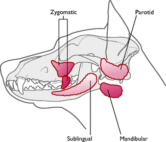
These are paired glands lying around the area of the oral cavity. Their secretions, known collectively as saliva, pour into the cavity via ducts. Saliva contains 99% water and 1% mucus – there are no enzymes in the saliva of the dog and the cat (Fig. 9.6).
The positions of the glands are:
< div class='tao-gold-member'>
Stay updated, free articles. Join our Telegram channel

Full access? Get Clinical Tree


