Chapter 19 Diencephalon
To understand the anatomic terminology used in this chapter, the following description is based on terms described in the Nomina Anatomica Veterinaria, fifth edition. The prosencephalon is the forebrain that consists of the telencephalon and diencephalon. The telencephalon is the endbrain, the principal components of which are the neopallium, rhinencephalon, and corpus striatum (basal nuclei) of the cerebrum, which consists of two cerebral hemispheres. The diencephalon is the intermediate brain that consists of the thalamencephalon, hypothalamus, and subthalamus. The thalamencephalon includes the thalamus, metathalamus, and epithalamus (Box 19-1).
DIENCEPHALON
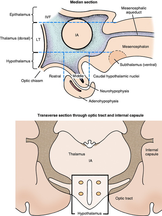
The components of the diencephalon are distributed symmetrically on both sides of the third ventricle. The hypothalamus consists of specific nuclei that are located lateral and ventral to the ventral portion of the third ventricle, ventral to the interthalamic adhesion (Fig. 19-1). The subthalamus is between the thalamus and substantia nigra of the mesencephalon. It is composed of the subthalamic body and zona incerta. The thalamus is composed of a plethora of nuclei that make up the major part of the diencephalon located dorsal to the ventral portion of the third ventricle and medial to the internal capsule. The metathalamus consists of the medial and lateral geniculate nuclei. The epithalamus includes the habenular nuclei and their connections and the pineal gland. For teaching purposes, this text considers the metathalamic nuclei to be part of the thalamus. This chapter describes the major components of the thalamus and hypothalamus. The epithalamus was previously considered with the limbic system and the subthalamus with the extrapyramidal system.
THALAMUS
Anatomy
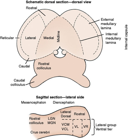
The thalamus is related to the hypothalamus ventrally and to the internal capsule laterally (see Fig. 19-1; see also Fig 2-3 through 2-5).7,29-32,35 Leptomeninges (pia and arachnoid trabeculations) and cerebrospinal fluid (CSF) cover most of its dorsal surface. It is composed of numerous nuclei partly separated by medullary laminae and arranged in a bilaterally symmetric pattern on either side of the third ventricle. The internal medullary lamina divides each side of the thalamus into medial and lateral halves, and it splits rostrally to enclose a rostral portion, which includes the rostral thalamic nucleus (Fig. 19-2). A dorsal plane through the lateral half separates the thalamus into dorsal and ventral tiers of nuclei, and two transverse planes through the ventral tier further define these nuclei from rostral to caudal. The thin external medullary lamina forms the external boundary of the lateral half of the thalamus and is separated from the internal capsule by a narrow nuclear area, the thalamic reticular nucleus. These divisions result in the following groups of nuclei with a listing of their specific nuclei.
Function
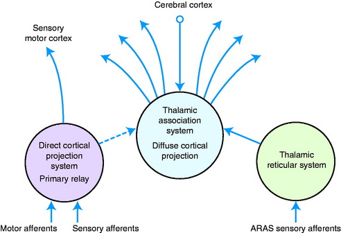
On a functional basis, the thalamic nuclei can be grouped into three major systems: (1) the direct cortical projection system, (2) the diffuse cortical projection system, and (3) the thalamic reticular system (Fig. 19-3).
Direct Cortical Projection System
The direct cortical projection system is the primary relay system that has been described throughout this text in relation to the conscious perception pathways for sensory systems and the thalamic relay for motor systems.
For the sensory systems, a thalamic relay occurs in the conscious perception pathway of all sensory systems except olfaction. The thalamic nuclei concerned with this relay are located in the ventral tier of the lateral half of the thalamus and in the caudal thalamic group. These thalamic nuclei are listed here with the specific sensory pathways afferent to the thalamic nucleus and the general area of the cerebrum to which each nucleus projects.
All of these primary relay nuclei also project to other thalamic nuclei.
Diffuse Cortical Projection System
The diffuse cortical projection system is the thalamic association system that receives axons only from other diencephalic nuclei and telencephalic sources. These sources include the primary relay thalamic nuclei and the thalamic reticular system nuclei, the hypothalamus, the cingulate gyrus, the frontal cortex, and the striatum. No afferents are received from the primary afferent pathways in the brainstem. These diffuse cortical projection nuclei project diffusely to all parts of the telencephalon (see Fig. 19-3).
Thalamic Reticular System
The thalamic reticular system is the most rostral component of the ARAS, which receives afferents from more caudal brainstem levels of the reticular formation and collaterals of all the conscious perception pathways for sensory systems. Efferents from this system project to the thalamic nuclei of the diffuse cortical projection system (thalamic association system), which, in turn, projects diffusely to the telencephalic cerebral cortex (see Fig. 19-3). Thalamic nuclei that comprise this system include nuclei in the intralaminar (midline) group and the thalamic reticular nucleus.
Experimental evidence for the efferent pathways and functions of these various thalamic systems comes from the following studies.26,28
Ascending Reticular Activating System
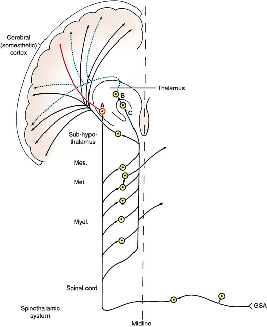
The reticular formation is defined in Chapter 8, and the ascending component, the ARAS, is introduced at that time. The ARAS is part of the reticular formation, which, you may recall, consists of a network of neurons in the central portion or core of the brainstem from the medulla through the pons and midbrain and into the diencephalon.21 The ARAS receives afferents from all the conscious projection pathways of the sensory systems, which includes interoception, exteroception, and proprioception. As these conscious projection pathways (spinal and cranial) course rostrally through the brainstem to the primary relay nuclei in the thalamus, collateral axons are given off into the reticular formation (Fig. 19-4). Most of these collaterals synapse in the reticular formation. Neurons of the reticular formation that project rostrally (the ARAS) continue the impulse flow rostrally in a multisynaptic pattern to one of two areas in the diencephalon, either the thalamic reticular system or the hyposubthalamic reticular system. A few collaterals project rostrally as part of the ARAS without synaptic interruption to terminate on one of these two diencephalic areas. The diencephalic portion of the ARAS stimulates the entire cerebral cortex diffusely. The thalamic reticular system affects the cerebral cortex by way of the diffuse telencephalic connections of the thalamic association system.
Summary of Thalamic Function
Stay updated, free articles. Join our Telegram channel

Full access? Get Clinical Tree


