CHAPTER 31 Diagnostic Imaging of the Ear
IMAGING OPTIONS
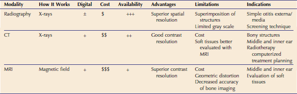
Sophistication of imaging in veterinary medicine continues to increase, following the advancements seen in human medicine. The imaging possibilities for evaluating the feline ear include radiography, CT, and MRI. Each of these modalities works differently and has advantages and disadvantages with regard to cost, availability, limitations, and indications (Table 31-1).
RADIOGRAPHY
Within radiography there are now two options for image capture. In all systems an x-ray tube is used to create the x-ray beam; the difference lies in the final display and generation of the image. The radiographic image can be captured with radiographic film that is developed either by hand-processing or an automatic processor. This is referred to as conventional film-screen radiography and is still used widely in veterinary practice. Digital radiography (DR) is beginning to replace conventional film-screen radiology in veterinary practice. Computed radiography (CR) is a form of DR. The x-ray beam interacts with the CR imaging plate and these imaging plates are processed by a plate reader, which creates a digital image on the computer. A computer image also is created with direct digital radiography (DDR), but there is no imaging plate to process. The DDR plate is incorporated into the radiographic table and sends the signal measured from the x-ray beam directly to a computer. With the increasing availability of digital radiography, computer manipulation and storage of radiographs are now possible. For more information on DR imaging, the reader is directed elsewhere.1
An important strength of radiography is the global information it can provide. In contrast, with cross-sectional imaging of CT and MRI, the anatomy is seen one “slice” at a time and the overall size and shape of organs must be reconstructed. Radiography is an excellent modality for bone imaging, which is what is evaluated in routine ear imaging (bullae, temporal bone). Radiography has high spatial resolution, which is the ability to discern small detail. The greatest disadvantage of radiography is the superimposition of overlying structures, which is at its greatest when imaging the skull. For example, it is not possible to image the brain with conventional radiography because of the overlying calvarium; the cross-sectional imaging modalities CT and MRI are more suitable for brain imaging. With conventional radiography it is not possible to discern whether the soft-tissue opacity within the bulla identified on radiographs is fluid or a mass because these two structures are the same opacity. However, CT and MRI can make that assessment more accurately, because these modalities have higher contrast resolution than radiography. Contrast resolution is the ability to discriminate between different tissues accurately and display these differences with varying shades of gray or brightness.2 Although conventional radiography has limitations, it still has its place in imaging of the feline ear, for example, in the assessment of otitis media.
COMPUTED TOMOGRAPHY
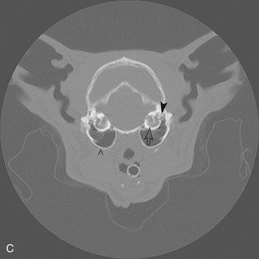
CT images are computer-generated information obtained from x-ray detectors and are based on differences in x-ray absorption within the patient, which result from variances in physical density between tissues. In CT a thin x-ray beam emitted from an x-ray tube is directed at the patient as it rotates around the animal. X-ray detectors opposite the x-ray tube measure the transmitted radiation, which is relayed to the computer for reconstruction into the tomographic image (Figure 31-1).
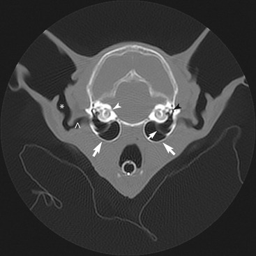
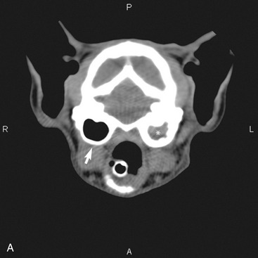
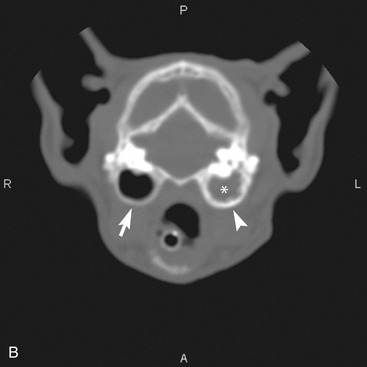
CT offers distinct advantages over conventional radiography. The images are tomographic, in other words “slices” of the patient. With tomographic images, superimposition of structures is not seen, which is a serious limitation of routine radiography. In CT imaging, contrast between structures is enhanced greatly because images are computer generated. There are only five radiographic opacities that we can identify on routine radiographs; and tissues, especially soft tissues, must be altered significantly in order to see a change. This is not true with CT images in which the range of tissue attenuation, or opacity, depiction is much greater (3000 to 4000 units), thereby increasing the accuracy of image interpretation. CT technology can display a larger gray scale than radiography, enhancing contrast between structures. The human eye can not visualize, and monitors can not display, the full range of gray scale on one CT image; therefore images must be windowed. This allows viewing of images with both a soft-tissue and a bone window by adjusting the gray scale on the monitor, and is necessary to evaluate the tympanic bullae accurately (Figure 31-2).3 Bone windows have window widths (WW) around 2000 Hounsfield units (HU) and window level (WL) of around 125 to 300, whereas a soft-tissue window will have a WW of 300 to 400 HU and a WL of 50 to 60 HU. A more in-depth discussion of CT image manipulation can be found elsewhere.2
Accurate detection of soft-tissue changes, delineation of tumors, and subtle bony lysis and proliferation are possible with the increased contrast afforded by CT. It has been shown that CT and radiographs have similar sensitivity for detection of otitis media.4 Therefore radiography of tympanic bullae still remains a cost-effective screening tool for ear disease. Although CT is increasingly available for our veterinary patients, it is more costly than routine radiographs and may not have a primary role as a first-line screening technique, being reserved for more complicated cases of ear disease such as neoplasia (see Chapter 69).
MAGNETIC RESONANCE IMAGING
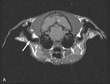
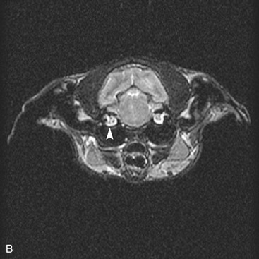
MRI is a tomographic imaging technique based on the properties of hydrogen atoms when placed in magnetic and radiofrequency fields. Hydrogen atoms are abundant in water and mammalian tissue, being highly prevalent in muscle and blood, and to a lesser extent in fat and bone. Because of this characteristic, MRI provides superior soft-tissue detail and is the imaging choice for the central nervous system. An in-depth discussion of the principles and equipment involved in image acquisition is beyond the scope of this chapter; additional information can be found elsewhere.2
Despite the superior soft-tissue detail obtained with MRI, there are instances when CT may be sufficient or a more desirable imaging modality. Because of the physical structure of bone, especially compact bone of the skull, the hydrogen atoms are not as movable and the bones of the skull appear black, an image void (Figure 31-3). Magnetic field inhomogeneity artifacts present with MRI can cause geometric distortion,5 limiting the accuracy of measurements. This is not encountered in CT imaging. Therefore because structures are more geometrically precise with CT, this is the preferred modality for computerized radiotherapy treatment planning.