Diagnostic Analgesia

Chapter 10 Diagnostic Analgesia


Despite the many technological advances in equine sports medicine over the past three decades, diagnostic analgesia arguably remains the most valuable tool in the equine clinician’s arsenal to localize pain causing lameness. Although the technique requires a thorough understanding of anatomy, basic technical skill, and clinical experience, the equipment and expense are minimal. In addition, diagnostic analgesia can be performed on site, with the outcome immediately obvious. Any lingering concern that a suspected “shoulder problem” exists is convincingly erased when the response to perineural analgesia of the digit is observed. This chapter reviews the various perineural, intrasynovial, and local (regional) infiltration techniques for application of local analgesia in the diagnosis of lameness in horses.


Local Anesthetics: Pharmacology and Tissue Interactions


Pain is transmitted specifically in the small, lightly myelinated, A delta and nonmyelinated C nerve fibers.1 All commonly used local anesthetic solutions, regardless of the specific molecular structure, share the same basic mechanism of action—specifically, the ability to block or inhibit nociceptive nerve conduction by preventing the increase in membrane permeability to sodium ions.2 These agents consist of a lipophilic and a hydrophilic group, connected by an intermediate chain containing a carbonyl group of an amide or ester linkage, and have traditionally been categorized as either amide- or ester-type local anesthetics.3 Common local anesthetic solutions used in horses—2% solutions of lidocaine, mepivacaine, and bupivacaine—are of the amide type.


Compared with most local anesthetics, lidocaine and mepivacaine are considered relatively fast-acting and have a reported duration of action of image to 3 hours and 2 to 3 hours, respectively. In contrast, bupivacaine is intermediate in onset but has a much longer duration of action (3 to 6 hours).4 Bupivacaine is most suited for providing therapeutic rather than diagnostic analgesia. The results in clinical practice vary, because in severely lame horses the degree and duration of local analgesia are decreased, regardless of the agent used.


When local anesthetic solutions are injected, tissue damage can occur but is extremely rare.3,4 Soft tissue swelling occurs occasionally and is likely caused by needle trauma or hematoma formation and not from a direct drug-tissue interaction. We suggest that alcohol and a clean wrap be applied to the injection sites when the diagnostic evaluation is complete to prevent or minimize swelling at injection sites. Cellulitis or other forms of infection are rare potential complications.


Acute synovitis, or flare, is a rare complication that can occur after intrasynovial (most commonly intraarticular) injection of local anesthetic solutions. Synovitis from intrasynovial injection of local anesthetic solution is much less common than from injection of other medications. Mepivacaine is thought to be less irritating than lidocaine when administered intraarticularly, but we have not recognized this difference.3 However, Dyson reported that lidocaine may be considerably more irritating than mepivacaine, and clinical data documenting differences were used successfully in the licensing of mepivacaine in the United Kingdom.5 Like cellulitis after perineural injections of local anesthetic solutions, infectious synovitis is a rare but possible sequela. To mitigate the possibility of contaminated solution, we use a new vial of local anesthetic solution when performing intrasynovial analgesic procedures.


Systemic side effects from diagnostic analgesic techniques are exceedingly rare. Cardiovascular or central nervous system signs, including muscle fasciculation, ataxia, and collapse, were reported.3 Systemic intoxication would require a dose much higher than is commonly used, even for an extensive diagnostic evaluation. For example, the maximum single infiltration dose of lidocaine that can be safely administered to a 500-kg horse is about 6.0 g, or 300 mL of a 2% solution.6


Strategy, Methodology, and Other Considerations


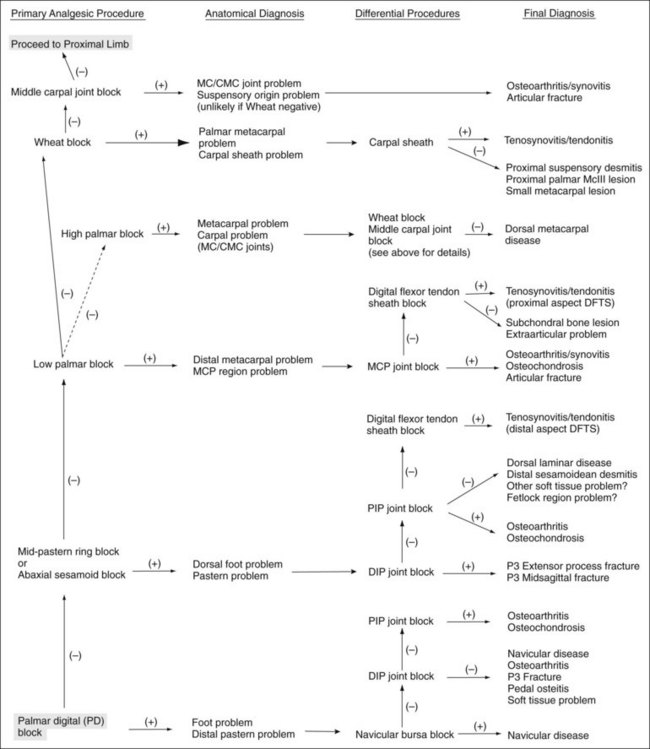
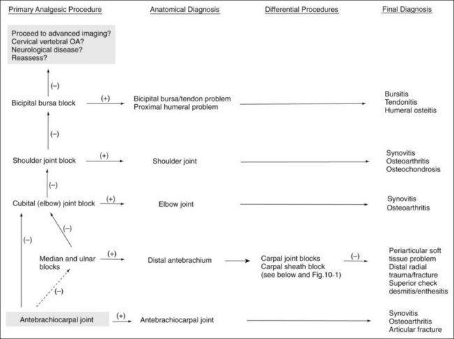
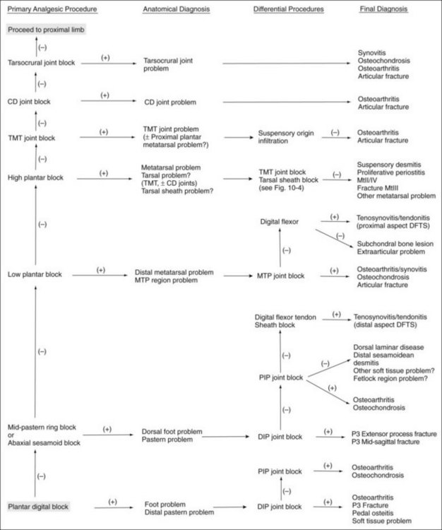
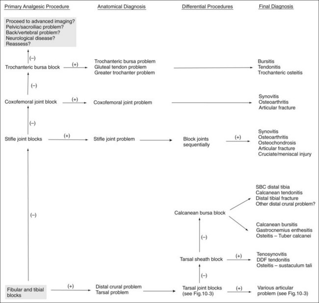
A few basic principles must be followed to ensure success. A thorough working knowledge of regional anatomy is required. Even for seasoned veterans a review of anatomy may be required before less common techniques are performed. A most important principle when performing perineural analgesia is to start distally in the limb and work proximally (Figures 10-1 to 10-4). If possible, sequential blocks from distal to proximal should always be used, but in certain circumstances a different strategy can be successful. Sequential blocking requires a fair amount of time, and in certain horses, selective intraarticular or local blocks can be performed without following this “golden rule.” However, in most situations, blocking a large portion of the distal limb at a proximally located site may preclude accurate determination of the source of pain causing lameness and may require an additional visit to perform additional diagnostic procedures.


It is important to test the efficacy of a perineural block before reevaluating the horse’s degree of lameness. If any question exists, the block should be repeated rather than assuming deep pain has been abolished, when skin sensitivity persists. If a horse shows partial improvement only minutes after injection, an additional few minutes should be allowed for complete analgesia to be achieved before proceeding with the next block. Alternatively, the block can be repeated. In so doing, the clinician minimizes the potential for misinterpretation and the tendency to ascribe the residual lameness to a “second problem” that does not exist.


During this portion of the examination, we are attempting to eliminate baseline rather than induced lameness, and care must be taken when adopting the practice of “blocking out a positive flexion test” (see Chapter 8). Once baseline lameness has been eliminated, we rarely perform additional flexion tests or attempt to eliminate all induced lameness.


How is the efficacy of the block assessed? Several methods are available, but the following points should be considered. Individual horses react differently to noxious stimuli applied to the skin. Therefore it is helpful to test the contralateral (unblocked) limb to establish the horse’s baseline response to the test. Similarly, covering a horse’s eye or feigning a few gestures with an instrument (pen tip, hemostatic forceps) without actually contacting the skin can help differentiate between a random or anticipatory response by an apprehensive horse and a true painful response. Positioning oneself on the contralateral side of the horse when testing for sensation also can help in making this determination. The clinician should avoid using sharp instruments that can penetrate the skin and cause hemorrhage, a situation not well understood by a concerned horse owner. Hemostatic forceps, used to pinch the skin, are ideal, because they are blunt and appear to consistently induce an appropriate amount of pain. Forceps are only useful in assessing superficial or skin sensation, however.


Perineural blocks must be assessed for the amelioration of deep and not just superficial pain. To assess whether deep pain in the hoof has been ameliorated after palmar digital analgesia or other techniques, hoof testers can usually be applied with enough force to cause a painful response, even in the most stoic of horses. Physical strength of the operator must be considered. Extreme or hard joint flexion (combined with varus or valgus stress) can be used to assess whether deep pain has been abolished in more proximal locations. In some instances, however, it is impossible to avoid contacting the skin proximal to the site of local anesthetic administration, leading the clinician to assume that the block has not worked. The application of firm digital pressure in the blocked area may be a viable alternative to flexion or manipulation to help avoid these potentially confounding factors.


It is important to understand that the region of the limb that is actually desensitized may, in fact, differ from the region the clinician intended to desensitize.7 Proximal diffusion of local anesthetic solution appears to be the most likely cause, but other, intangible factors may play a role. Using a small volume of local anesthetic solution (1 to 5 mL for most perineural blocks) can minimize but not abolish this phenomenon. To further minimize the potential for diffusion of local anesthetic solution, the horse should be reevaluated no more than 10 minutes after the injection (exceptions apply in certain situations). A recent study using 2 mL of radiopaque contrast medium injected perineurally around the palmar nerves at the level of the proximal sesamoid bones demonstrated proximal diffusion extending 2 to 3 cm within 10 minutes of injection, irrespective of whether horses stood still or walked.8


Complete analgesia, and thus 100% improvement in lameness score, is the goal when performing diagnostic analgesia, but in many horses this level of pain relief is never achieved. Improvement in degree of lameness greater than 70% to 80% after most perineural or intraarticular techniques should be considered a positive response in most horses. The quintessential response is that the horse “switches lameness” to the contralateral limb, indicating that now pain arising from the opposite limb is greater than the pain that caused the baseline lameness. However, complete response may not occur, and the clinician must decide when to stop sequential blocks or when the horse has “blocked out.” The clinician hopes for an obvious difference in lameness score when the horse is blocked, but in some horses, serial improvement occurs with each successive block, a situation that makes assessing the primary source of pain difficult.


Incomplete response to local analgesia in some horses may be explained by the fact that chronic pain, particularly deep bone pain, may remain resistant to complete analgesia when perineural techniques are used. For example, horses with laminitis tend to remain lame despite blocking many times at the appropriate level, probably because of neuropathic pain.9 Mechanical gait deficits do not improve after diagnostic analgesia because pain is minimal. Horses may continue to show lameness even with pain abolition, a situation that appears to be caused by habit. These horses tend to show mild residual lameness initially, only to warm out of it quickly during examination. Other factors affecting response to diagnostic analgesia include individual variation in neuroanatomy, the intermittent nature of certain lameness conditions, and the inherent difficulty in assessing and abolishing pain in horses with subtle lameness.10 Articular and subchondral bone lesions may not be desensitized by intraarticular analgesia, and pain may be more effectively abolished using perineural techniques.


Sensory innervation of joints is complex and involves three classes of neurons that transmit information from four receptor types, each of which has a specific distribution throughout the joint.11-13 Articular pain can arise from several sources, including the synovium (inflammation, effusion), fibrous joint capsule (increased intraarticular pressure), articular and periarticular ligaments, periosteum, and subchondral bone (injury, osseous vascular engorgement).10,12,14,15 Other than small branches in the perichondrium, articular cartilage is devoid of innervation. In osteoarthritic joints, however, erosion channels, formed in the calcified layer of cartilage, are invaded by subchondral vasculature.12 Putative nociceptive neurotransmitters were identified in these areas, and therefore it is plausible that in horses with advanced osteoarthritis, pain could be emanating from the deep cartilage layers.16,17


On occasion, lameness from an articular lesion abates after perineural analgesia but shows minimal or only partial response after intraarticular analgesia. In some horses, this can be explained by the fact that pain is originating from articular and periarticular tissues.8 Subchondral bone pain—caused by maladaptive bone remodeling, cystic or erosive lesions, incomplete fractures, and osteoarthritis—is inconsistently abolished by intraarticular analgesia. In fact, subchondral bone pain is abolished much more consistently by perineural techniques. Subchondral bone receives innervation from endosteal branches of peripheral nerves that enter the medullary cavity through the nutrient foramen.10,11,18,19 Intraarticularly administered local anesthetic solutions may not penetrate subchondral bone sufficiently to completely block these nerves. This shortcoming is presumably even more likely in situations in which the cartilage is intact.


Unfortunately, intraarticular analgesia, although easier to perform, inconsistently abolishes pain from many of the common articular problems. This fact, however, is either overlooked or misunderstood by many practitioners. Whenever possible, perineural analgesia should be performed, particularly in the distal aspect of the limbs, because this type of analgesia more consistently abolishes pain from all aspects of the joint and surrounding soft tissue structures.


Lameness Is Worse after Diagnostic Analgesia


Two uncommon situations arise when performing diagnostic analgesia. The first occurs during a blocking session. After performing palmar digital analgesia in a horse with forelimb lameness, lameness score may worsen by one or two grades. In fact, lameness may occasionally be considerably worse, prompting concern by the lameness diagnostician. Why? This unusual response occurs most commonly in horses with proximal, palmar metacarpal pain caused by proximal suspensory desmitis, avulsion fracture of the proximal palmar aspect of the third metacarpal bone (McIII), or proximally located superficial digital flexor tendonitis. Horses normally shorten the cranial phase of the stride to protect a source of pain, a common response by any lame horse. We reason that after palmar digital analgesia a lack of proprioception in the digit prompts the horse to take a somewhat longer stride, increasing the cranial phase. Compared with before the block an exaggerated load causes the horse to display a higher lameness score. Temporary exacerbation of lameness after palmar digital analgesia can be a useful characteristic to help determine the genuine source of pain.


The second situation is more ominous. A horse will occasionally be very lame, sometimes non–weight bearing, once a block wears off. This unusual, but important and sometimes difficult situation occurs when incomplete fractures become separated, displaced, or comminuted. Horses with fractures of the distal phalanx that are incomplete or have healed partially by a fibrous union or those with incomplete fractures of the proximal phalanx appear most at risk. Horses at risk are candidates for imaging before blocking, but in some this complication is unforeseen (see following discussion).


Horse Preparation


Before perineural analgesia is performed, the skin and hair should be cleaned of any gross debris such as mud, bedding, feces, or poultice. Clipping usually is not necessary unless the hair coat is long and prohibits accurate palpation of anatomical landmarks or adequate cleaning of the site. The site should then be scrubbed with an antiseptic, such as povidone-iodine or chlorhexidine, using clean gauze sponges or cotton. If the clinician has any concern about inadvertent penetration of a synovial cavity, a 5-minute aseptic preparation should be performed. This is followed by isopropyl alcohol administration over the site using cotton or gauze sponges.


Aseptic preparation should always be performed before any intrasynovial injection. Considerable debate and variation exists among clinicians regarding the need to clip the hair over the site. Some clinicians always clip the hair, whereas others never do. Still others shave the hair in a small area directly over the injection site. The results of a study indicated no significant difference in the number of postscrub colony-forming units (bacterial flora) between clipped and unclipped skin over the distal interphalangeal (DIP) and carpal joints.20 Nonetheless, we still clip the hair over all proposed intrasynovial injection sites before undertaking a 5-minute aseptic preparation. The only time we deviate from this policy is when we are specifically asked not to clip the hair, a situation that arises in some sports horses actively competing, in claiming horses, or in those being sold.


Similar variation among clinicians exists regarding wearing of sterile latex gloves when performing an intrasynovial injection. However, we recommend wearing sterile gloves during these procedures. Science aside, clipping hair and wearing sterile gloves project a positive impression to all in attendance.


How does, or should, the practitioner attempt perineural or intrasynovial analgesia in a horse with contact or chemical dermatitis (scurf) over the proposed injection site? A superficial wound or abrasion with a localized infection presents a similar quandary. For obvious reasons, these areas are difficult, if not impossible, to clean effectively. If possible, an alternative site, away from the area of dermatitis, should be used. If not, then the procedure should be delayed until the skin condition (or wound) has resolved. In many instances, dermatitis can be treated with topical medications (medicated sweats such as nitrofurazone-dimethylsulfoxide) for a few days to facilitate resolution of the problem.


Injection Techniques


Perineural injections are typically performed using needles ranging in size from 25 gauge, 1.6 cm (image inch) to 20 gauge, 2.5 or 4 cm (1 to image inches). Small needles cause less pain but carry the risk of breaking off within tissues if the horse kicks out or otherwise misbehaves. For this reason, we recommend using 18- or 19-gauge needles for injections or blocks within the proximal metatarsal or plantar tarsal regions. In the distal aspect of the limb the needle is inserted subcutaneously directly over and parallel to the nerve. We generally direct the needle proximally rather than distally, although this portion of the procedure differs among clinicians. One of the Editors (SJD) always inserts the needle distally; if a fractious horse throws the limb to the ground the needle is more likely to stay in situ, and the remainder of the procedure may be completed with the limb on the ground. Directing the needle distally also ensures more distal placement of the local anesthetic solution, which may be important at distal sites. The needle is inserted before the syringe is attached. To avoid excessive manipulation once the needle is inserted, a slip-type syringe hub is preferred. Syringes with screw-on hubs can be difficult to attach, requiring additional manipulation in a sometimes fractious horse, and are not generally used. However, when dense tissue requires that additional force be used for injection, the seal between the hub and the needle can be broken, a complication minimized by using a screw-on hub (see the following discussion of lateral palmar block).


Volume of local anesthetic solution varies, but for a majority of blocks in the distal limb, 1 to 5 mL is injected at each site. Larger volumes are used to perform the median and ulnar or fibular (peroneal) and tibial techniques and when infiltrating the proximal palmar (plantar) metacarpal (metatarsal) region. After injection, we briefly massage the sites with gauze sponges or clean cotton soaked in alcohol. Skin sensation and deep pain are assessed 5 to 10 minutes after injection. More time is allowed under certain circumstances (see specific comments throughout the chapter). At the completion of the examination an alcohol wrap should be applied to minimize swelling, a common sequela resulting from local irritation and bleeding from nearby vessels.


For “ring” blocks, circumferential subcutaneous infiltration of local anesthetic solution, and other local or regional infiltration techniques, we most commonly use 20- to 22-gauge, 4-cm needles. For performance of a ring block, the needle is inserted perpendicular to the long axis of the limb, and local anesthetic solution is injected as the needle is advanced, leaving a clearly visible wheal or subcutaneous bleb in most locations. The needle then is reinserted at the leading edge of this wheal, a practice that minimizes the number of injections and the horse’s discomfort. However, most horses object to needle insertion even when it is performed well within the bleb. The injection is continued around the limb in this manner. For most ring blocks in the distal limb, 10 to 15 mL of local anesthetic solution is used, but larger volumes may be preferred for surgical procedures. Ring blocks can be done as a substitute for or in combination with perineural injections (see the specific blocking techniques discussed in the chapter). However, simply placing local anesthetic solution in a subcutaneous location is not a substitute for the preferred approach, direct perineural injection.


To block a local area such as a splint or curb, the needle is typically inserted in one or two locations, and local anesthetic solution is deposited in a fan-shaped pattern. As with the perineural analgesia, the sites are massaged briefly and the horse is reevaluated in 5 to 10 minutes.


Intrasynovial injections typically are performed using needles ranging in size from 22 gauge, 2.5 cm to 18 gauge, 4 cm. If marked effusion is present, drainage of synovial fluid is advised, either by allowing the fluid to drip from the hub of the needle or by aspirating with a sterile syringe before proceeding with injection. We prefer the former procedure unless fluid analysis is necessary. The manipulation required to attach the syringe may cause the horse discomfort and potentially dislodge the needle but if successful may hasten withdrawal of synovial fluid. Brief evaluation of the color and viscosity of synovial fluid can shed some light on the disease process within and is expected practice among most racehorse trainers. Volume of local anesthetic solution varies considerably between synovial cavities, but the clinician should keep in mind that small volumes might contribute to a false-negative result. False-negative results are common in horses with severe osteoarthritis, and larger volumes of local anesthetic solution should be used. We routinely spray or wipe antiseptic solution over the injection site. After the examination a light bandage is applied over the injection sites from the metacarpophalangeal or metatarsophalangeal joint, distally. Initial reevaluation is done 5 to 10 minutes after injection. Additional evaluations may be necessary depending on the response during the initial time period. General practice is to have the horse walked in hand or with a rider after perineural or intrasynovial analgesia is administered, a procedure thought to hasten distribution of local anesthetic solution and potentially improve success. Excessive diffusion of local anesthetic solution is a potential drawback to this practice, particularly with techniques such as DIP or middle carpal analgesia (see the following discussion), although it would be a complication difficult to quantify.


Another issue to consider when performing diagnostic analgesia is whether riding or driving a horse after blocks have been performed is safe. In general, riding on the flat or driving a horse at slow speed after any of the common blocks have been performed is safe. Stumbling or knuckling can be a concern after upper limb perineural techniques, such as the median and ulnar and fibular (peroneal) and tibial techniques. Common sense should prevail, however, with regard to the horse and rider negotiating fences or performing at high speed. Horses at risk for lameness from stress or incomplete fracture are candidates for imaging before evaluation at speed after diagnostic analgesic techniques have been performed. Moreover, horses suspected of having incomplete fractures but with negative or equivocal radiological findings may best be managed conservatively without use of analgesic techniques and should undergo either follow-up radiographic examination in 10 to 14 days or scintigraphic examination.


Perineural Analgesia in the Forelimb


Palmar Digital Analgesia


Palmar digital analgesia (or palmar digital block) is the most common diagnostic analgesic procedure performed. The medial and lateral digital neurovascular bundles, consisting, in a dorsal to palmar direction, of the digital vein, artery, and nerve, course in an abaxial location to the digital flexor tendons. With the exception of small breeds or draft horses with remarkably long-haired pasterns (feathers), the palmar digital nerve is easily palpable between the proximal sesamoid bones and the cartilages of the foot. The palmar digital block can be performed with the horse in a standing position or with the limb held off the ground. We prefer the latter. If held by an assistant, the limb should be grasped in the midmetacarpal region, with the fetlock and digit hanging in neutral position. The palmar digital nerve is easily palpated in this extended position on the lateral aspect of the deep digital flexor tendon (DDFT). Alternatively, the clinician performing the block can hold the limb, a technique that requires practice. The clinician can stand facing backward with a hand grasping the midpastern region or can stand behind the limb and clutch the hoof between both legs.


A 25-gauge, 1.6-cm needle is inserted subcutaneously, directly over the nerve, just proximal to the cartilages of the foot (Figure 10-5). One of us (LHB) directs the needle in a distal direction, whereas the other (MWR) directs the needle in a proximal direction to avoid deeper penetration or laceration of digital vessels if the horse withdraws the limb. Alternatively, a 22-gauge, 4-cm needle can be inserted on the palmar midline in the midpastern region, and local anesthetic solution is then infiltrated in a V-shaped pattern. This modification of the palmar digital block is quite difficult to perform in the hindlimb but when done in the forelimb provides maximal analgesia to the bulbs of the heel and minimizes the potential for depositing local anesthetic solution dorsal to the nerve. Loss of skin sensation in the midline between the bulbs of the heels should be assessed, because this area seems most recalcitrant to palmar digital analgesia. Deep pain is assessed using hoof testers. However, if skin sensation persists, it is still worth reevaluating lameness, because in some horses deep pain and lameness may be abolished despite the persistence of skin sensation.


Traditionally the palmar digital block was felt consistently to desensitize only the palmar (plantar) one third to one half of the foot.21 However, in clinical practice, this block desensitizes 70% to 80% of the foot. Most of the DIP joint is affected, with the exception of the proximodorsal aspect. Horses with fractures of the extensor process of the distal phalanx or injury of a collateral ligament of the DIP joint may show partial improvement after palmar digital analgesia, however. Our clinical observations have been substantiated in a recent study. Setscrews were placed near the medial and lateral aspects of the toe to simulate pain from the sole. Lameness in these horses was abolished using palmar digital analgesia performed just proximal to the heel bulbs.22


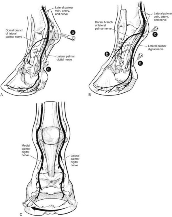
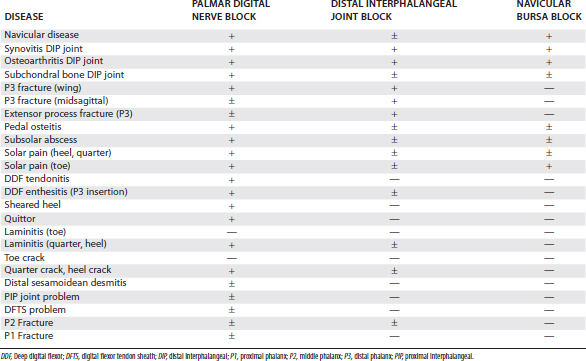
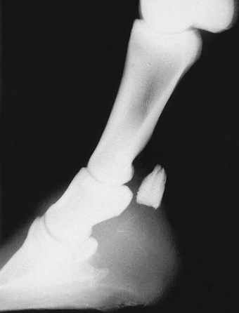
Classically, most horses that responded positively to palmar digital analgesia were thought to have navicular syndrome, but this block desensitizes many lameness conditions within and outside the hoof capsule (Table 10-1). This is an important and common misconception. Lameness in horses with proximal interphalangeal joint pain, midsagittal fracture of the proximal phalanx, or other conditions involving the fetlock joints can be abolished using palmar digital analgesia.7,23 Although using small volumes of local anesthetic solution and performing the block just above the cartilages of the foot may help to minimize the area of analgesia, these procedures do not prevent inadvertent diagnosis in some horses. Diffusion of local anesthetic solution is the most likely explanation, and even a small volume can readily spread in a proximal direction, but the normal anatomy of the digit prevents distal placement of local anesthetic solution (Figure 10-6).


The concept that palmar digital analgesia abolishes lameness in an area considerably more than the palmar (plantar) one third of the foot appears to be difficult for many to accept. Although results of studies are widely published and this finding has been emphasized at international meetings, most veterinary students still graduate today armed with this common misconception. Diffusion of local anesthetic solution easily explains why lameness conditions in the proximal aspect of the pastern or fetlock regions are desensitized by palmar digital analgesia. But what about the innervation of the hoof itself? Skeptics should consider the anatomy of the palmar digital nerve. Most practitioners have severed the palmar digital nerve while performing neurectomy. Can the clinicians recall any instance of having identified a large dorsal branch, or for that matter, any branching of the nerve at all? The lack of nerve branches in the midpastern region is circumstantial evidence that important innervation to the structures located dorsally within the hoof capsule occurs farther proximally (ill-defined dorsal branches) or after the nerve courses deep to the cartilages of the foot. It makes little sense that ill-defined dorsal branches would innervate the dorsal two thirds of the foot, leaving the robust palmar digital nerve to innervate only the palmar one third. When carefully dissected the palmar digital nerves can be seen branching extensively deep to the cartilages of the foot, sending branches dorsally to innervate the dorsal portions of the foot.


Accurately quantifying the contribution of the palmar digital nerve to the innervation of the foot or, for that matter, the exact percentage of structures desensitized by palmar digital analgesia may be impossible. Clinical experience will undoubtedly convince practitioners of the broad nature of palmar digital analgesia. Finally, it is imperative to develop expertise in diagnostic imaging of the entire digit, because the many lameness conditions affected by palmar digital analgesia require detective-like differential diagnostic skills.


Midpastern Ring Block


Traditionally the diagnostic blocks performed after palmar digital analgesia are the basisesamoid or abaxial sesamoid techniques. The basisesamoid block provides little additional information compared with palmar digital analgesia, unless, of course, the dorsal branch, originating from the digital nerve at the level of the proximal sesamoid bones, is blocked. If, however, the dorsal branch is blocked, then the basisesamoid block is in reality an abaxial sesamoid block. For this reason, we rarely perform the basisesamoid block. When performing the abaxial sesamoid technique in racehorses, or, for that matter, in any sport horse with a propensity to develop lameness of the metacarpophalangeal or metatarsophalangeal joints, the veterinarian runs the risk of an additional misdiagnosis. When local anesthetic solution is deposited in a location abaxial to the proximal sesamoid bones, pain from the metacarpophalangeal or metatarsophalangeal joints can be inadvertently blocked, explained most likely because of diffusion of local anesthetic solution, leading the clinician to assume the horse has a problem in the foot or digit, but in reality the pain originated from these joints. For these reasons, we prefer to use a blocking sequence as follows: palmar digital nerve, followed by a dorsally directed subcutaneous ring block, followed by the low palmar or plantar block.


The midpastern ring block affects the dorsal branches of the digital nerves and desensitizes any remaining areas of the foot and pastern region that were not affected by palmar digital analgesia. In most horses this includes the dorsal 20% of the foot (dorsal laminar and extensor process regions of the distal phalanx) and the dorsal pastern region (dorsal aspects of the middle phalanx and proximal interphalangeal joint, and distal portions of the proximal phalanx). Although desirable, performing the dorsal ring block just above the cartilages of the foot usually is not possible. Instead the block is performed at the level of the midpastern region.


A 20- to 22-gauge, 4-cm needle is used to deposit subcutaneously 10 to 12 mL of local anesthetic solution, beginning near the injection site used for palmar digital analgesia over the lateral neurovascular bundle and continuing dorsally and medially, ending over the medial neurovascular bundle (see Figure 10-5). Resistance to needle advancement and injection of local anesthetic solution will invariably be encountered dorsally, if the block is done just proximal to the coronary band, because of the dense tissue (proximal interphalangeal joint capsule, extensor branches of the suspensory ligament, and extensor tendons). Performing the block in the midpastern region minimizes this problem and mitigates the potential for inadvertent penetration of the proximal interphalangeal joint.


Abaxial Sesamoid Block


Desensitizing the medial and lateral palmar nerves at the level of the proximal sesamoid bones is commonly referred to as the abaxial sesamoid block but may provide the same information as the basisesamoid block, if the dorsal branch of the palmar digital nerve is blocked. To avoid redundancy, we rarely perform the basisesamoid technique before progressing to the abaxial sesamoid block (see previous comments). A block done at this level essentially provides analgesia of the entire digit, because the block is performed at the level of or just proximal to the origin of the dorsal branch of the palmar digital nerve. Response to this block may vary, however. Some horses retain skin sensation in the dorsoproximal aspect of the pastern region. In others, pain arising from lesions involving the fetlock joint or periarticular tissues is abolished. In part, these phenomena can be explained by proximal diffusion of local anesthetic solution, affecting the palmar digital nerves proximal to the fetlock joint. Branches of the palmar digital nerves supplying the proximal sesamoid bones, the sesamoidean nerves, could easily be blocked using an analgesic technique in this abaxial position.24 One of the Editors (SJD) regularly performs palmar nerve blocks at the base of the proximal sesamoid bones as a first block if, on the basis of clinical examination, it is considered unlikely that the horse has foot-related pain and there is also no evidence of likely fetlock joint pain.


The abaxial sesamoid block can be performed in the standing horse or with the limb held by the clinician or an assistant. The assistant grasps the foot, facing forward. The assistant should be warned that a fractious horse may kick backwards with the limb, so he or she should stand slightly to one side, outside the plane of the limb. The palmar digital nerve can easily be palpated over the rigid proximal sesamoid bones and in fact is in its most superficial position in this location. A 25-gauge, 1.6-cm needle is directed in a proximal or distal direction and typically 1 to 3 mL of local anesthetic solution are used for each of the medial and lateral injections. Deep pain is assessed by hard flexion of the interphalangeal joints. False-negative or delayed results can arise because of deposition of local anesthetic solution outside the fascia that surrounds the neurovascular bundle.8


Low Palmar Analgesia


Analgesia of the metacarpophalangeal joint region and distal aspect of the limb is induced using the low palmar block or low palmar analgesia (low four-point). This technique blocks the medial and lateral palmar nerves and the medial and lateral palmar metacarpal nerves. In the forelimb a subcutaneous, dorsally directed ring block and block of the dorsal branch of the ulnar nerve completely abolishes skin sensation. Disagreement exists about whether abolishing skin sensation is necessary when performing perineural techniques. Abolition of skin sensation independently from nerves contributing to deep pain sensation, as in the case of the low palmar technique, does not necessarily mean deep pain is abolished, which is particularly relevant when a nerve responsible for skin sensation is blocked. When using these techniques for diagnostic purposes, it may be best to avoid blocking nerves that contribute only skin sensation, thus minimizing the number of needle insertions For therapeutic interventions, however, these nerves need to be blocked.


The low palmar block is performed at the level of the distal end (bell or button) of the second and fourth metacarpal bones (splint bones), with the limb in a standing position or held off the ground (Figure 10-7). A 20- or 22-gauge needle is used to inject 1.5 to 5 mL of local anesthetic solution at each injection site. To block the palmar metacarpal nerves, the needle is inserted perpendicular to the skin, just distal to the end of the splint bones, to a depth of 1 to 2 cm. It is important to deposit local anesthetic solution deep in the injection site, rather than simply in a subcutaneous location. While local anesthetic solution is continuously injected, the needle is slowly withdrawn, leaving a visible bleb in the subcutaneous space. To block the medial and lateral palmar nerves, the needle is inserted subcutaneously, in the palmar aspect of the space between the suspensory ligament and DDFT at the level of or slightly more proximal to the distal end of the splint bone. To improve the accuracy of the injection, using a fan-shaped injection technique is helpful. If the digital flexor tendon sheath (DFTS) is distended, the injections must be performed more proximally. Inadvertent penetration of the DFTS is possible even if it is not distended, so careful skin preparation is mandatory. To complete this block, local anesthetic solution is placed in the subcutaneous tissues from the bleb at the distal end of the splint bone to the dorsal midline. One of the Editors (SJD) does not do this last step.


An alternative technique to abolish pain associated with maladaptive or nonadaptive bone remodeling or other causes of subchondral bone pain of the distal aspect of the McIII is to block the lateral and medial palmar metacarpal nerves separately from the lateral and medial palmar nerves. In some horses suspected of having this injury, use of abbreviated low palmar analgesia will avoid additional injections of local anesthetic solution. With this technique the lateral or medial palmar metacarpal nerve, or both, can be blocked individually or together, and the horse’s gait assessed. In many horses with this cause of lameness, contralateral forelimb lameness will then be seen. If lameness does not abate, the clinician then completes low palmar analgesia using the technique described previously (see the following discussion).


Alternatively, some clinicians prefer to use a longer needle first to deposit local anesthetic solution over the palmar metacarpal (metatarsal) nerves. The needle is then pushed subcutaneously to deposit local anesthetic solution over the palmar nerves (see Figure 10-7). When this modification is performed, incompletely blocking the palmar metacarpal (metatarsal) nerves or lacerating the digital vessels is possible. The lateral and medial palmar nerves can be blocked using only the lateral injection site by advancing the needle in a medial direction, palmar to the DDFT. Although each of these modifications may theoretically decrease the number of injections needed to perform this technique, they have the disadvantages of potential hemorrhage and incomplete analgesia.
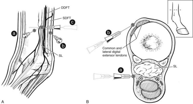


High Palmar Block


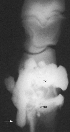
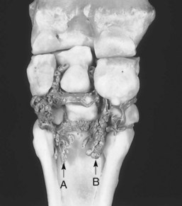
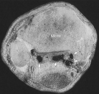
To provide analgesia to the metacarpal region, the high palmar block (high four-point, subcarpal block) is the most common technique, but a modified block (lateral palmar or Wheat block) can be performed. Inadvertent penetration of the carpometacarpal joint is a potential complication with the high palmar block. A similar complication can occur in the hindlimb but is less frequent (see the following discussion). Inadvertent penetration of the carpometacarpal joint occurred in 17% of specimens, in which a conventional high palmar block was performed, because of extensive distopalmar outpouchings (Figures 10-8 and 10-9). However, when the high palmar block was performed within 2.5 cm of the carpometacarpal joint, inadvertent penetration of this joint occurred in 67% of specimens. The carpometacarpal joint always communicates with the middle carpal joint, and therefore penetration of the carpometacarpal joint during high palmar analgesia would lead the clinician to diagnose a metacarpal problem, when in reality the authentic lameness condition exists in the carpus. Moving the injection site in a distal direction decreases the possibility of entering the carpometacarpal joint but also narrows the scope of the technique and may result in a false-negative response in a horse with proximal suspensory desmitis. Two ways around this likely complication are these: first, the clinician could perform middle carpal analgesia before performing high palmar analgesia; second, the clinician could perform a lateral palmar block in lieu of the conventional high palmar technique. In an experimental study, the carpal joints were unlikely to be entered inadvertently during performance of the lateral palmar block, although in every specimen, local anesthetic solution would have entered the carpal canal.25 Unless the clinician is familiar with the lateral palmar block, the most straightforward approach to reduce the possibility of misdiagnosis in this region is to perform middle carpal analgesia before proceeding to the high palmar block. When local anesthetic solution is placed in the middle carpal joint, not only is the carpometacarpal joint blocked, but also the possibility exists of providing local analgesia to the proximal palmar metacarpal region. With this approach, abolishing pain associated with proximal suspensory attachment avulsion injury (desmitis, fracture), stress remodeling, and longitudinal fracture is possible (see Chapter 37). The palmar metacarpal nerves and suspensory branches from the lateral palmar nerve are closely associated with the distopalmar outpouchings of the carpometacarpal joint, and diffusion of local anesthetic solution from this area could explain in part this clinical finding (Figure 10-10).


It is important for the clinician to understand that interpretation of analgesic techniques in the proximal palmar metacarpal region or carpus can be somewhat complex. Correct diagnosis is always the key, and comprehensive evaluation using multiple imaging modalities is a must in differentiating lameness in this region. From the clinical perspective, one is more likely to assume incorrectly that one is dealing with a carpal problem when the authentic lameness condition resides in the proximal palmar metacarpal region than vice versa. Numerous techniques are used to perform high palmar analgesia; some provide partial and others provide complete analgesia to the metacarpal region. For complete analgesia, blocking the following nerves is necessary: the medial and lateral palmar nerves, the medial and lateral palmar metacarpal nerves, the suspensory branches, and nerves providing skin sensation along the dorsum (dorsal branch of ulnar nerve and musculocutaneous nerve). To block these nerves effectively, one must use a site close to the carpometacarpal joint, at the level where the splint bones begin to taper (Figure 10-11

Only gold members can continue reading. Log In or Register to continue

Stay updated, free articles. Join our Telegram channel

Jun 4, 2016 | Posted by in EQUINE MEDICINE | Comments Off on Diagnostic Analgesia

Full access? Get Clinical Tree

Get Clinical Tree app for offline access