chapter 9 Developing a Technique Chart
Upon completion of this chapter, the reader should be able to do the following:
Santes’ rule: Calculation for determining an approximate amount of kilovoltage (kVp) necessary for a given anatomic area on the basis of measurement and the grid being used: (2 × tissue thickness in cm) + source-image distance + grid factor = kVp.
Technique chart: A chart based on tissue thickness and anatomic part that can be consulted for predetermined machine settings.
INTRODUCTION
The technique chart is an invaluable resource for a radiographer. Its purpose is to provide a consistent method of choosing the proper exposure factors to create a diagnostic radiograph. On the basis of the thickness of tissue and anatomic area of the body, the radiographer can consult the technique chart for a predetermined machine setting. Without this resource, one would have to calculate a new technique each time a radiograph is taken or use a questionable technique performed previously. A technique chart prevents the need for second radiographs due to inappropriate exposure factors.
Every x-ray machine should have its own formulated technique chart. It is often thought that a successful exposure technique used on x-ray machine A will also work on x-ray machine B. This is not true. Even x-ray machines of the same make and model vary in both quantity and quality of output because of variations in input voltage and calibration. Several other factors influence the chart:
The factors that affect a technique chart should be standardized as much as possible. The SID, amount of beam filtration, film processing, and type of grid should remain constant. The success of a technique chart depends on the radiographer’s willingness to maintain continuity of the variables.
The only variables that should change are the types of film and screens used. The film type and speed should be preselected and limited. The screen speed chosen should fit the needs of the practice and be limited to one or two speeds. A veterinary practice that has a number of screen types must formulate many technique charts. Working with a number of film and screen types that have coinciding technique charts can be confusing and may increase errors.
SUGGESTED CHARTS
Another misconception is that just one technique chart is necessary for an x-ray machine. On the contrary, anatomic and technical differences call for more than one chart. Several charts may be needed and may include the following:
In general, five (screen) variable kVp technique charts based on species and anatomy are recommended for a small-animal veterinary practice:
TECHNIQUE CHART FORMULATION
Several methods are used in veterinary practice to formulate a technique chart; they vary slightly, but all are effective. The method presented here is different from others because of a few shortcuts that the technologist may find helpful. The principles of technique chart formulation can be applied to any species and anatomic area. The method presented here applies to x-ray machines that have variable kVp and exposure time settings.
To create a workable technique chart, a series of trial exposures must be made using a cooperative, average-sized patient. Theoretically, by exposing one radiograph, all five suggested technique charts can be formulated.
Equipment needed for this procedure should be gathered before proceeding. A mature dog with average conformation (neither obese nor emaciated) and weighing approximately 50 pounds is an ideal patient for this procedure. With this size dog, a 14- × 17-inch cassette should be used. Use medium-speed x-ray film because of its versatility in veterinary practice.
Exposure Factors
To formulate a variable kVp technique chart, a test radiograph is made of the canine abdomen in lateral recumbency. The measurement is in the range of 11 to 16 cm, and the exposure factors are based on the screen type and grid ratio used. Remember, any measurement exceeding 10 cm necessitates the use of a grid to reduce fog-producing scatter radiation. For an SID of 40 inches, the following information applies:
Base mAs Factors
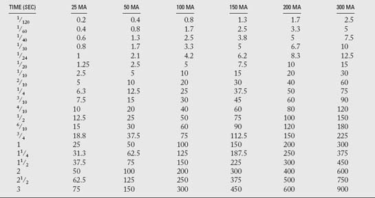
An mAs chart is shown in Table 9-1. The following base mAs requirements for the intensifying screens are merely starting points for the radiographer. Each radiographic system may require slightly different exposures.
Screen Type | mAs |
---|---|
Fast (high speed) | 2.5 to 10 |
Medium (par speed) | 5 to 12.5 |
Slow (ultradetail) | 30 to 40 |
Exposure Modification for Grid Use
When using a grid, increased exposure is necessary to maintain adequate radiographic density. The addition of a grid usually requires doubling the exposure time. For example, if the exposure technique needed for a tabletop (nongrid) exposure is 2.5 mAs (1/120 second at 300 mA), the new exposure for grid use would be at least 5 mAs (1/60 second at 300 mA).
The kVp will also need to be modified. The amount of modification varies according to the grid ratio being used.

Stay updated, free articles. Join our Telegram channel

Full access? Get Clinical Tree

