Curb

Chapter 78Curb


The historical definition of curb is enlargement on the plantar aspect of the fibular tarsal bone (calcaneus) caused by inflammation and thickening of the (long) plantar ligament.1 There is confusion regarding the cause of curb; a seminal publication uses this historical definition in the text but describes curb as superficial digital flexor (SDF) tendonitis in a figure legend.2 By ultrasonographic evaluation curb was redefined as a complex of soft tissue injuries that occurs on the distal plantar aspect of the tarsus.3,4 Long plantar (LP) desmitis is only one of many injuries that causes curb. However, the indelible term curb has been used for hundreds of years5 and is still useful to describe swelling of the distal, plantar aspect of the tarsus (excluding the calcaneal bursa and proximal aspect of the calcaneus). In the rest of this chapter, curb is used specifically to mean the clinically apparent soft tissue swelling of the plantar aspect of the tarsus seen best from the side. Conformational abnormalities or bony exostoses can mimic or contribute eventually to formation of curb.


Clinical Appearance of Curb


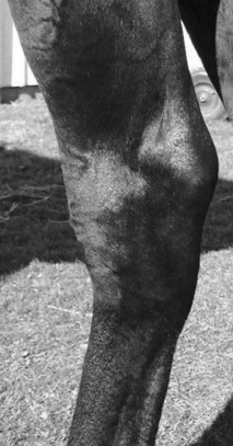
The convex profile typical of curb is best seen from the side (Figure 78-1). Careful evaluation of swelling from all perspectives, palpation, and thorough lameness examination are critical. Curb must be differentiated from other swellings of the hock, including capped hock, effusion, edema and fibrosis of the calcaneal bursa (see Chapter 79), tarsal tenosynovitis (see Chapter 76), thoroughpin (with or without involvement of the tarsal sheath), and bony enlargements of the distal hock region (see Chapter 44). Injuries of the deep digital flexor tendon (DDFT) as it courses along the plantaromedial aspect of the hock within the tarsal sheath can produce typical signs of curb, but they account for only a small percentage of injuries in horses with curb.


Horses with sickle-hocked conformation are said to be curby (see Chapter 4). Sickle-hocked and in-at-the-hock conformation lead directly to curb, a finding most common in Standardbred (STB) and Thoroughbred (TB) racehorses. Prognosis for STB racehorses with sickle-hocked conformation and curb is worse in a trotter than in a pacer. Trotters with sickle-hocked conformation are usually fast early in training and racing, but this conformation is often career limiting. Sickle-hocked conformation is also undesirable in TB racehorses. Horses with sickle-hocked conformation often develop curb first, but they then independently or concomitantly develop other lameness associated with the distal hock joints. Tarsal region lameness begins with curb in 2- and 3-year-olds and progresses to osteoarthritis of the centrodistal and tarsometatarsal joints or slab fractures of the central tarsal bone, or more commonly the third tarsal bone.


Horses can have curby conformation without developing curb, and horses with normal hindlimb conformation can develop curb. The proximal aspect of the fourth metatarsal bone (MtIV) is often prominent in horses with sickle-hocked conformation. The most dramatic example of altered joint morphology occurs in young foals with tarsal crush syndrome, the result of delayed or incomplete ossification of the tarsal cuboidal bones (see Chapter 44).


Firm, fibrous soft tissue swelling can develop just proximal to the MtIV as a sequela to injection or local analgesia of the tarsometatarsal joint, presumably from local trauma, hemorrhage, or leakage into the subcutaneous tissues. Mild bony proliferation or fragmentation of the proximal aspect of the MtIV, or of the fourth tarsal bone, can cause focal swelling easily mistaken for curb.


Considerable variation in clinical signs occurs, and the injury cannot be categorized, or a management program and prognosis established, without thorough clinical and ultrasonographic examinations. Historically, owners and trainers consider curb to be an annoying, self-limiting problem that rarely causes lameness or poor performance, that responds to a single treatment that is uniformly effective, and that is cured by treatment. Most racehorse trainers are opposed to resting a horse with curb unless lameness is performance limiting, so veterinarians often are faced with management decisions without an option for even short-term rest or a reduction in training intensity. Many traditional therapies have no data to support efficacy. Thus, progress in understanding and management of this complex soft tissue injury has been limited.


Lameness ranges from none, mild, or severe depending on the structure involved and extent of injury. Lameness tends to be worse if the soft tissue structure involved is located dorsal to the superficial digital flexor tendon (SDFT) in the plantar aspect of the tarsus (i.e., the DDFT and/or the long plantar ligament [LPL]), if SDFT injury is diffuse, or if a mixed injury involves more than one structure. Diagnosis is straightforward in horses with obvious lameness seen at a trot in hand and painful swelling, but lameness may be evident only as a slight loss of performance or unlevelness when the horse is performing maximally and may be perceived only by trainers, drivers, or riders. A horse with chronic curb may not exhibit signs of pain during palpation, or lameness at a trot in hand, but can show lameness at speed, and convincing a trainer that the long-term swelling is a source of pain may be difficult. The area should be palpated carefully with the limb bearing weight and flexed. Swelling may be firm and fibrous, with few signs of active inflammation, or may be warm, painful, and edematous. Acute, compliant, or mushy swelling is associated with hemorrhage or other subcutaneous fluid accumulation and sometimes deeper soft tissue injury. Horses with this form of curb usually have acute, moderate-to-severe lameness. Horses with distal hock joint pain often exhibit a painful response when direct pressure is placed on plantar hock structures, including the SDFT, proximal aspect of the MtIV, second metatarsal bone, and proximal aspect of the suspensory ligament. Often swelling is not detected in these horses. Response to upper limb flexion varies and is nonspecific. Direct digital palpation followed by trotting is useful, because horses with active inflammation show increased lameness.


Because horses with curb can have concomitant osteoarthritis or other problems of the distal hock joints, differentiation of the source of pain is important but difficult. Diagnostic analgesia is useful but not foolproof. If horses are lame at a trot in hand, local infiltration of local anesthetic solution subcutaneously over the curb is effective. A minimum of 20 to 30 mL of local anesthetic solution should be infiltrated along the lateral, plantar, and medial aspects of the curb. Small-gauge needles should be avoided (to avoid needle breakage), and the injection should be performed with the limb in flexion, because horses may object to several injections. If horses are not visibly lame at a trot in hand, examination at the track or under saddle should be performed. Selective intraarticular analgesia of the distal hock joints and sequential perineural analgesia to rule out the lower limb are essential. A tibial nerve block alleviates pain with curb, but it is seldom done because other common sources of pain are abolished similarly.


Radiography and scintigraphy help differentiate other sources of pain, but ultrasonography is the imaging method of choice to determine which structures are involved and the extent of damage.


Applied Anatomy and Normal Ultrasonographic Examination of the Plantar Aspect of the Tarsus


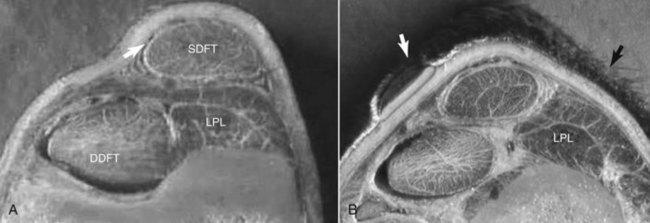
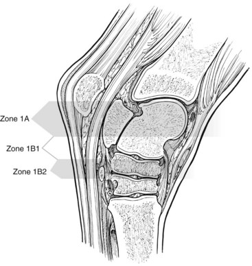
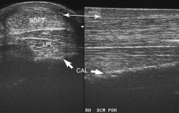
Plantar to the calcaneus are skin, subcutaneous tissues, a thin fibrous tissue layer, the SDFT, and the LPL (Figure 78-2). Medially the DDFT courses distally over the sustentaculum tali, within the tarsal sheath. Normally the tarsal sheath has a small amount of fluid that can be seen during ultrasonographic examination, but it is not felt. The LPL originates from the calcaneus, closely adheres to this bone, and inserts distally on the plantar surface of the fourth tarsal bone and the MtIV. The plantar aspect of the tarsus can be divided into zones to classify findings (Figure 78-3) or the distance measured from the proximal aspect of the calcaneus (point of hock). Swelling comprising curb occurs in zone 1 of the tarsal and metatarsal regions but can extend into zone 2 if SDF tendonitis occurs. Transverse and longitudinal images of both limbs should be obtained from the plantar midline, plantaromedial (to evaluate the DDFT), and slightly plantarolateral (to evaluate the distal aspect of the LPL). Measurement of cross-sectional area (CSA) is important to confirm lesions in which enlargement has occurred but with no overt fiber damage. Precise placement of the ultrasound transducer is important because the LPL changes size and shape as it courses distally. Knowledge of normal ultrasonographic anatomy is crucial (Figures 78-4 to 78-7).3,4


Jun 4, 2016 | Posted by in EQUINE MEDICINE | Comments Off on Curb

Full access? Get Clinical Tree

Get Clinical Tree app for offline access