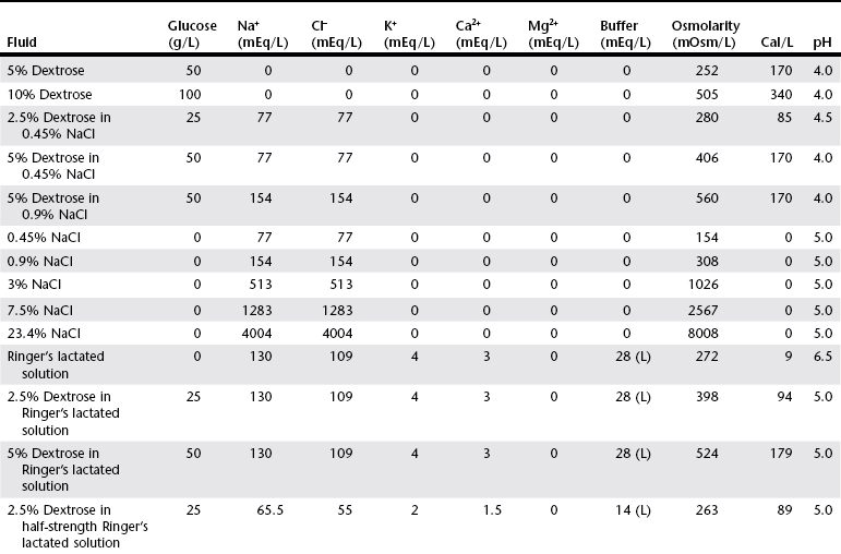
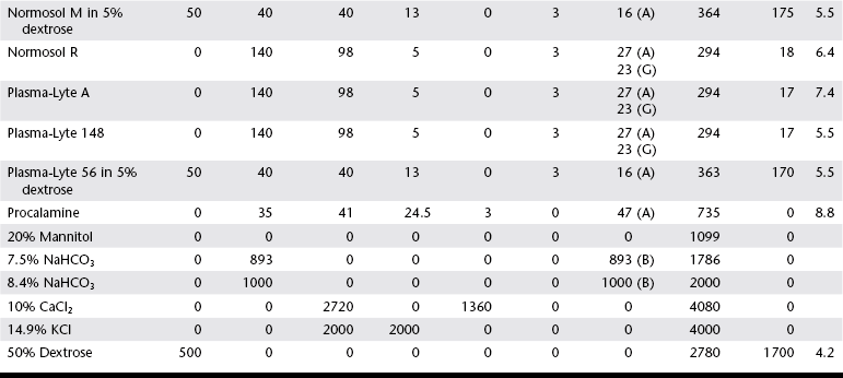
Crystalloid Fluid Therapy

Chapter 1


Crystalloid Fluid Therapy


Crystalloids are water-based solutions containing electrolyte and nonelectrolyte solutes and are capable of entering all body compartments. They are the most common fluid type used therapeutically in veterinary medicine.


The major goals of crystalloid fluid therapy are restoration of intravascular volume (in shock), replacement of interstitial fluid deficits (in dehydration), and provision of maintenance fluid needs for dogs or cats at risk of dehydration. Fluid therapy is not warranted in cases in which heart failure cannot be excluded, as in the hypothermic tachypneic cat, and it should be used with caution if anuria is a possibility. The route of fluid administration is determined based on the goals of fluid therapy and the likelihood that these goals may be reasonably met with the planned therapeutic approach. The authors assume that most practitioners employ fluid therapy on a regular basis, and thus the purpose of this chapter is to highlight some controversial and emerging aspects of fluid therapy.


Types of Crystalloid Fluids


Crystalloids are classified as isotonic, hypotonic, or hypertonic in relation to plasma osmolality. Isotonic crystalloids are by far the most commonly used fluid type in veterinary medicine (Table 1-1). Also known as replacement fluids, isotonic crystalloids are used to replace fluid deficits that may have developed from excessive loss. The electrolyte composition of isotonic fluid is typically similar to that of plasma. These solutions may also be classified as acidifying (e.g., normal saline) or buffering (e.g., lactated Ringer’s solution [LRS], Normosol-R, Plasma-Lyte 148) solutions. Buffering solutions are considered balanced due to an electrolyte composition that is roughly equal to plasma. Normal saline is not a balanced solution and is acidifying due to a relatively high concentration of chloride and a lack of buffer, both of which decrease plasma bicarbonate.


The clinical relevance of isotonic fluid choice is perhaps less important than commonly believed, although it is reasonable to closely evaluate the electrolyte composition of the fluid choice in relation to the patient’s needs. For example, 0.9% saline might be preferred in cases of head trauma, hypercalcemia, or metabolic alkalosis caused by gastrointestinal (GI) obstruction.


Some believe that LRS administration to patients with hepatic dysfunction is contraindicated. A recent study in healthy dogs found that LRS given at a rate of 180 ml/kg/hr resulted in a significant increase in plasma lactate concentration within 10 minutes of infusion. However, the clinical significance of this finding is uncertain, especially considering the extreme fluid rates needed to raise the plasma lactate. Additionally, the impact of lactate-containing solutions has never been investigated in patients with liver disease. In the absence of fulminant low-output hepatic failure, the benefits of using LRS for treatment of hypovolemia and metabolic acidosis likely outweigh the risks. Alternatively, balanced isotonic crystalloids with acetate or gluconate may be considered. However, these molecules also require hepatic metabolism to bicarbonate (albeit less than LRS) to exert their buffering effects.


Hypotonic crystalloids have a lower tonicity relative to plasma and contain additional free water. These solutions may be termed maintenance fluids. Hypotonic fluids are primarily used when volume replacement is adequate but ongoing fluid therapy is deemed necessary, such as for a dog recovering from maxillary fracture that is not yet eating. When fluid therapy is needed in the patient with cardiac failure, hypotonic solutions also may be preferable.


Hypertonic crystalloids (e.g., 7.5% saline) can be used for short-term, rapid resuscitation and intravascular volume expansion, as well as for treatment of head trauma. These solutions pull water from the interstitium down the concentration gradient into the intravascular space. They are preferred in euhydrated but hypovolemic patients. These fluids cause interstitial and intracellular dehydration and therefore must be followed by administration of isotonic crystalloids. The addition of electrolytes and dextrose may increase the tonicity of isotonic fluids to a hyperosmolar range (see Table 1-1). Hypertonic crystalloids are not intended for long-term use.


Fluid Management of Hypovolemic Shock


Hypovolemic shock can occur in dogs and cats in association with trauma, massive gastrointestinal hemorrhage, spontaneous hemoperitoneum (e.g., ruptured splenic mass), untreated gastrointestinal disease (e.g., parvovirus infection), gastric dilation-volvulus (GDV), diabetic ketoacidosis, and renal failure. Fluid administration is a mainstay of shock management.


The term shock dose of fluids has been popularized in emergency and critical care medicine. Twenty years ago it was commonly recommended to give a specific dose of fluids to animals in shock (generally 90 ml/kg in dogs and 60 ml/kg in cats). The volume was set to replace an entire blood volume with crystalloids administered over a period of 1 hour or less. Similarly, administration of shock volumes of fluids was considered the norm in human patients until publication of the landmark study investigating immediate versus delayed fluid resuscitation for hypotensive patients (Bickell et al, 1994). The results of that study challenged the idea that if some fluids are good, more must be better. Multiple experimental studies and clinical trials in people have subsequently demonstrated that excessive crystalloid or synthetic colloid fluid therapy is associated with dilutional coagulopathy, decreased wound healing, increased risk of sepsis, and acute lung injury leading to acute respiratory distress syndrome (ALI/ARDS). Current resuscitation efforts in massively traumatized humans have focused on treatment with blood and blood products. It is unlikely that most veterinary hospitals have a blood bank with sufficient resources to resuscitate all dogs and cats with blood, but this may become a therapeutic possibility in the future.


So what do these studies mean for the clinician treating a patient in hypovolemic shock? Intravascular fluid support is clearly indicated to restore or maintain circulating blood volume. However, the dose of fluid should not be a specific volume but rather a quantity titrated to restore perfusion parameters such as heart rate, mucous membrane color, and peripheral pulse quality to normal values. Blood pressure (BP) is maintained in most cases of shock owing to compensatory mechanisms. Normal BP does not exclude hypovolemia and should be interpreted with caution in patients with other signs of shock. Lactate is a useful surrogate marker of inadequate tissue perfusion, with increased lactate (>2.5 mmol/L) associated most commonly with hypoperfusion. It should be recognized that blood lactate may increase with patient struggling, with excessive muscle activity, in association with metabolic alternations (e.g., neoplasia), and with some medications, most notably prednisone (Boysen et al, 2009).


A reasonable starting point for fluid administration is 15 to 20 ml/kg of crystalloids over 10 to 15 minutes with reassessment of patient parameters, especially those of perfusion. Additionally, attention should be directed to the underlying cause of the hypovolemic shock so that the cause can be treated directly through surgical or other medical therapy. If a patient is not responding rapidly to initial fluid therapy, urgent response is needed to reverse shock. It helps to know the average response time for a specific disease and to understand other comorbidities that may accompany the specific injury. For example, if stabilization is not occurring, major differentials include ongoing bleeding into the abdomen, thorax, or fracture site (femur/pelvis) or possibly pneumothorax.


Other useful monitoring parameters include heart rate, respiratory rate, pulse quality, lactate, packed cell volume, total solids, urine production, and attitude. Goals for fluid therapy in shock are restoration of circulating plasma volume to reverse oxygen debt and normalization of the above parameters. Fluid therapy may be tapered over several hours if the resuscitation goals have been met and the patient remains stable.


Fluids should be given as a bolus in the patient with evidence of intravascular volume depletion, as suggested by tachycardia, dull mentation, pale mucous membranes, or hypotension. If vomiting and diarrhea are associated with extreme hemoconcentration, such as a packed cell volume of greater than 60% in non-greyhound dogs (or > 70% in greyhounds), a fluid bolus also may be used to decrease blood viscosity and improve hemodynamics. A fluid bolus is not needed in a pet that is simply dehydrated.


Hypotensive or Low-Volume Resuscitation


Extrapolating from medicine, limited-volume resuscitation (also know as low-volume or hypotensive resuscitation) has gained some momentum in veterinary medicine. The goal of low-volume resuscitation is administration of fluid volumes sufficient to achieve a mean arterial BP of 60 to 80 mm Hg, ensuring adequate tissue and organ perfusion while preventing clot disruption. This method, used for people with uncontrolled cavitary hemorrhage requiring surgical stabilization, aims to have patients in the operating room within an hour of first response to minimize the time that they are hypotensive. Short transitions from the emergency room to the operating room can be difficult to attain in the veterinary setting. Although surgical intervention is usually unnecessary for traumatic hemoabdomen, other causes of hemoabdomen, such as ruptured neoplasia, are likely to require surgery. In this latter setting, low-volume resuscitation may be beneficial for the patient earmarked for surgery. However, this approach requires careful patient monitoring so that prolonged hypotension and the complications of poor tissue perfusion and organ dysfunction are avoided.


Occasionally, there is confusion between low-volume resuscitation and resuscitation with hypertonic saline (HTS) and colloids. One popular method is to mix 43 ml of hetastarch with 17 ml of 23.4% hypertonic saline in a 60-ml syringe and then dose this at 4 to 5 ml/kg over 10 to 15 minutes. Although HTS and colloids represent a smaller volume of fluid than an equivalent dose of crystalloids and may be infused more rapidly than an isotonic crystalloid, the approaches are not interchangeable. Use of HTS and colloid results in a higher plasma volume than administered due to an interstitial fluid shift to the vascular space.


In one small unpublished study of nontraumatic hemoabdomen, dogs randomized to hypertonic saline and hetastarch showed a more rapid time to stabilization than did dogs randomized to crystalloids; however, no differences were noted in either outcome or transfusion needs.*

< div class='tao-gold-member'>

Only gold members can continue reading. Log In or Register to continue

Stay updated, free articles. Join our Telegram channel

Jul 18, 2016 | Posted by in PHARMACOLOGY, TOXICOLOGY & THERAPEUTICS | Comments Off on Crystalloid Fluid Therapy

Full access? Get Clinical Tree

Get Clinical Tree app for offline access