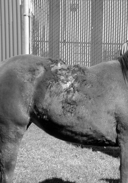
Stephen D. White In horses, albinism must be distinguished from lethal white syndrome, which is primarily a problem in Paint Horses (especially, but not exclusively, in overo breedings). The defective gene has also been found in American Miniature Horses, half-Arabians, Thoroughbreds, and crop-out Quarter Horses (foals born to registered Quarter Horses that have too much white to register with the American Quarter Horse Association). The Veterinary Genetics Laboratory at the School of Veterinary Medicine, University of California, Davis (VGL-UCD)1 offers a diagnostic test to determine carrier status. The lethality of albinism in horses comes from the association with intestinal aganglionosis. Affected foals die shortly after birth. Because some white foals are not affected, euthanasia should be performed only after signs of intestinal malfunction occur. This disease is similar to Hirschsprung’s disease in humans and is linked to a mutation in the endothelin-B receptor gene. Hereditary equine regional dermal asthenia (HERDA) develops early in life in horses. Most affected horses are Quarter Horses, but registered Paint Horses and Appaloosas with Quarter Horse lineage have developed this disease. Many of the Quarter Horses are from high-quality cutting horse lines. Although reported cases have been from North America, the disease and its associated genetic defect have been reported from Europe and South America. The disease (or similar condition) has also been reported in a crossbred Arabian mare, a Thoroughbred gelding, a Hanoverian foal, and a Haflinger horse. The working hypothesis for HERDA in horses is that there is a defect in the structure or healing process of the collagen fibers in the middle to deep part of the dermis. Affected horses have less cutaneous tensile strength in the skin than healthy horses. Typically, these areas are over the back and sides of the neck. The skin in these areas may be easily torn or stretched and often develops seromas and hematomas (“blisters” filled with serum or blood; Figure 134-1). Healing is usually adequate but often leaves unsightly scars. Diagnosis is often based on clinical signs alone; histologic findings are sometimes subtle, but clumped or poorly organized collagen fibers below the level of the hair follicles may be seen. A zone of mid-dermal to deep dermal separation has been reported in two horses and is observed in some biopsy samples. Poorly oriented collagen fibers are sometimes seen on electron microscopy. Interestingly, affected horses may also have decreased thickness of the corneas, increased tear production, and increased incidence of corneal ulcers. This condition is almost certainly present at birth, but HERDA is often not noticed until horses are about 2 years old, when training with tack and a saddle begins, and the resulting friction and trauma induce the typical lesions. As with many genetic diseases, no effective treatment or cure exists; some of these horses have been maintained as so-called pasture pets. This disease follows an autosomal recessive mode of inheritance, so in order for the foal to be affected, both the sire and the dam must carry the gene. If the same pair were bred again, there would be an approximately 25% chance that the next foal would also be affected. A genetic marker determined for this disease relates to a missense mutation in equine cyclophilin B (PPIB), which in recent work was seen to cause a functional defect in this protein, resulting in less effective catalysis of the rate-limiting step in collagen folding. The VGL-UCD offers a diagnostic test to determine carrier or affected status. Both carriers and clinically affected horses with HERDA should be removed from breeding programs. Epidermolysis bullosa (EB) includes a number of diseases typified in humans and animals by the common finding of blister formation after minor trauma. Most forms are congenital and become apparent soon after birth. In animals and humans, subsets of EB are classified by the histologic location of the blister or cleft. These subtypes and respective cleft location are termed EB simplex (involving the basal cell layer of epidermis), junctional EB (intralamina lucida or basal cell layer), and dystrophic EB (sublamina densa). Junctional EB has been reported in Belgian foals of both sexes, in other breeds, and in a donkey. Lesions are usually seen within 3 days of birth and include multiple asymmetrical skin erosions and ulcers, often encrusted. Lesions may be especially prominent around the coronary bands (causing the hoof to crack and slough) and on the oral, anal, and genital mucosa. Histology and ultrastructural findings indicate a cleft in the intralamina lucida zone of the basement membrane. This is presumably caused by a defect in the anchoring filaments that connect the basement membrane to filaments in the superficial dermis. A laminin-5 (laminin 3A32) defect has been demonstrated in Belgians and in two French draft breeds, Trait Breton and Trait Comtois; the mutation is a cytosine insertion in exon 10 of the LAMC2 gene. A diagnostic test to determine carrier status in Belgian draft horses and related breeds is available.2 Clinical presentation and age of an affected foal form the basis for a strong suspicion of EB. Histology and, ideally, electron microscopy are required to confirm the diagnosis. There is no known treatment, and affected horses, as well as the sires and dams of affected horses, should not be bred; the mode of inheritance is autosomal recessive. The disease previously termed epitheliogenesis imperfecta in American Saddlebreds is now recognized as junctional epidermolysis bullosa. Lesions are most common on the limbs, head, and tongue. Hooves may slough in severe cases. The clinical presentation is usually diagnostic. In moderately to severely affected animals, the disease is fatal within a few days; the foals die of septicemia or other developmental abnormalities. Mildly affected areas may heal by scar formation.
Congenital Skin Disorders
Lethal White Syndrome
Hereditary Equine Regional Dermal Asthenia (Hyperelastosis Cutis)
Epidermolysis Bullosa
Congenital Skin Disorders
Chapter 134
Only gold members can continue reading. Log In or Register to continue

Full access? Get Clinical Tree


