Conformation and Lameness

Chapter 4 Conformation and Lameness


The thought that the way a horse is conformed determines the way it moves is well accepted. The relationship of conformation, especially of the distal extremities, and lameness also is well recognized. “Conformation determines the shape, wear, flight of the foot, and distribution of weight.”2 Veterinarians often are asked to comment on conformation during lameness and prepurchase examinations, especially with regard to the suitability of the horse to perform the intended task. In some instances, as in the case of presale yearling evaluations, the veterinarian’s opinion is paramount, and purchase is contingent on judgment of the yearling’s potential to perform as a racehorse, given its conformation, or in some instances its conformational faults (see Chapter 99). “It is by a study of conformation that we assign to a horse the particular place and purpose to which he is best adapted as a living machine and estimate his capacity for work, and the highest success in this connection will be best attained by the judicious blending of practice with science.”3 Evaluation of conformation and its influence on lameness is based largely on observation, experience, and pattern recognition. Recognizing desirable conformational traits in horses suited for a particular sporting activity and learning when to overlook a minor fault that has little clinical relevance are important.


Hereditary Aspects of Conformation


Certain conformational faults appear to be highly heritable traits. Evaluation of broodmares and foals often reveals that the early conformational defects seen in a foal are present in the dam. The dam seems to contribute more to faulty conformation than does the sire, although the stallion also is important. This difference may be explained in part by the fact that fillies with faulty conformation may develop problems or be retired early and subsequently bred, whereas most stallions usually are proven performers with exceptional conformation. Conformational faults such as toed in and toed out commonly are passed down from generation to generation. Back-at-the-knee (calf-knee), offset (bench) knee, tied-in below the knee, sickle-hocked, and straight-behind conditions appear to be highly heritable. In a recent study abnormal sire phenotype (offset carpi and outward rotation) was associated with faulty yearling carpal conformation.4 Heavier foals and yearlings were more likely to have faulty carpal conformation and inward rotation of the fetlock joints.4


Certain lameness conditions are common in horses with faulty conformation, but similar lameness conditions develop inexplicably in some breeding lines year after year in offspring with apparently acceptable conformation. Lameness of the carpus or tarsus appears to be most important. For example, in Standardbreds (STBs), siblings commonly develop similar lameness conditions, such as proximal suspensory avulsion injury, carpal osteochondral fragments, distal hock joint pain, or curb.


Objective Evaluation of Conformation: Is It Possible?


An attempt to quantify conformation using a linear assessment trait evaluation system that allows the observer to assess where, given a particular trait, a single horse falls within a population of horses, was described in 1996.5 A population of 101 Irish Thoroughbred (TB) flat racehorses and 19 top stallions was used and 27 common conformational traits were evaluated, including various heights, lengths and angles, and distal extremity conformation. Of the 27 traits, six were significantly linked to age (withers’ height and conformation, back length, neck size, carpal conformation, and hind pastern conformation), and five were linked to sex (head and neck shape; neck size at the poll, the larynx the withers, and the manubrium of the sternum; and forelimb hoof pastern axis). Most traits exhibited large phenotypic variation within the population, but 21 of 27 non–age-linked traits were judged suitable for possible inclusion in a linear assessment protocol.4 Researchers judged a high percentage of horses to be toed out, suggesting this trait may even be desirable.


More recent studies have used video-image analysis, but direct physical measurements may be more accurate than those obtained by analyzing videotapes or photographs.6 Potential errors in image analysis occur because of movement of skin markers over selected bony protuberances, a phenomenon more common in the upper limb and in motion studies.6,7 Skin marker location is critical for evaluation of joint angulation and movement during locomotion or conformation analysis. Instantaneous center (or axis) of rotation (ICR) is defined as the point with zero velocity during movement of that joint; accurate measurements of joint angulation require positioning markers at the ICR.8 Conventional positions of skin markers and ICR in most joints agree well, but use of traditional marker sites on the scapulohumeral and femorotibial joints results in overestimation and underestimation, respectively, of caudal joint angles.9 Although video-image analysis may be fraught with potential or in some instances real error, objectivity is a major advantage. Other advantages include the ability to replay images, reduction of observer fatigue, elimination of observations and measurements in real time, and permanent recording of the observation. Ideally, objective measurements would withstand statistical evaluation, distinguish between desirable and undesirable traits, and account for differences among different types of horses.6


A combination of direct measurement and photography was used to evaluate conformation of Swedish Warmblood (WBL) and elite sports horses.10,11 Whereas most of the conformational defects were mild or moderate, 80% of WBL horses were toed out behind, suggesting this may be a normal finding in this breed as in the STB trotter. More than 50% of horses had bench knees and 5% were toed out in front, contrary to findings in STB trotters.10,12 Many of the elite horses were bucked kneed, whereas the riding school horses tended to have calf-knees. It was speculated that this occurred because elite horses were evaluated after competition, and muscle fatigue may have contributed to the tendency to be over at the knee. Sex had a significant influence on conformation; females were smaller and had longer bodies and smaller forearms and metacarpal regions. There were interesting findings regarding hock angle. A sickle hock is defined as a hock angle of 53 degrees or less; a large hock angle is referred to as straight behind. Sickle-hocked conformation was nearly absent in elite horses, and it was hypothesized that sickle-hocked conformation either predisposed a horse to lameness or impaired a horse’s ability to achieve upper levels of competition.10 A positive relationship between larger hock angles and soundness in STB trotters also exists.13 All results must be viewed in moderation, because it is our clinical impression that horses with excessively large hock angles (straight behind) are substantially predisposed to suspensory desmitis. In forelimbs of WBL show jumpers and the forelimbs and hindlimbs of STB trotters, smaller fetlock joint angles (less upright) were desirable.10,13


Radiology was used to assess the degree of hyperextension of the carpus to study the potential effect of back-at-the-knee (calf-knee) conformation on the subsequent development of carpal chip fractures.14 Lateromedial radiographic images of 21 horses with carpal chip fractures and of 10 normal horses were obtained, with and without the contralateral limb raised. No relationship between measured carpal angle and carpal chip fracture formation existed, suggesting that this group of TB racehorses did not develop carpal chip fractures as a result of calf-kneed conformation. The sample size was small, however, and a larger study may produce different results. Horses with severe calf-kneed conformation may develop other problems and not advance enough in training to develop carpal chip fractures. They may be judged poor surgical candidates, are not referred, or are slow.


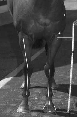
Two recent studies evaluated TBs and Quarter Horses (QHs) using skin markers, photography (three views: front, side, back), and computer-image analysis.15,16 Of the two studies done in TBs, one evaluated longitudinal development of conformation from weaning to 3 years of age. A strong relationship between long bone lengths and withers heights for all ages supported the theory that horses are proportional. Longitudinal bone growth in the distal limb increased only 5% to 7% and was presumably completed before the yearling year. Withers height, croup height, and length of neck topline, neck bottom line, scapula, humerus, radius, and femur increased significantly from age 0 to 1 year and age 1 to 2 years. Hoof lengths (medial and lateral, right and left) grew significantly from the ages of 0 to 1 and 1 to 2 years but decreased in length from age 2 to 3 years (presumably associated with trimming).15 Changes in growth measures indicated that growth rate either slowed or reached a plateau at 2 to 3 years of age. Horses also became more offset in the right forelimb between weaning and age 3, but the offset ratios did not change with age in the left forelimb. Shoulder angle increased in all age groups (becoming more upright), and this contributed to the increase in measured height at the withers. Dorsal hoof angle (both front and hind) decreased significantly from ages 0 to 1 and 1 to 2 years but did not change in the 2- and 3-year-old groups. This study provided objective information regarding conformation and skeletal growth in the TB, which could potentially be used for selection and recognition of important conformational abnormalities.15 Measurements of length and angle were obtained from photographs in which a tape measure was used for objective criteria and an objective method was developed for measuring offset knees (Figures 4-1 to 4-4).15
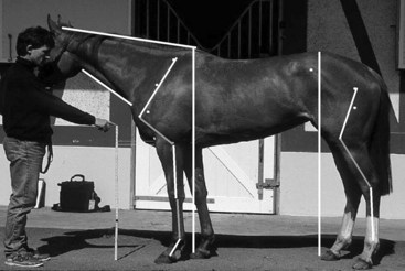
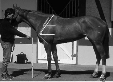


In another study the role of conformation in the development of musculoskeletal problems in the racing TB was evaluated.16 Conformation measurements were obtained from photographs of horses with markers at specific reference points and digitally analyzed as previously described.15 Clinical observations were recorded regularly for each horse, and stepwise (forward) logistic regression analysis was performed to investigate the relationship between binary response of clinical outcomes probability and conformation variables by the method of maximum likelihood. Clinical outcomes significantly (P < .05) associated with conformational variables included effusion of the front fetlock joints, effusion of the right carpal joint, effusion of the carpal joints, effusion of the hind fetlock joints, fractures of the left or right carpus, and right front fetlock and left hind fetlock lameness. Offset knees contributed to fetlock lameness (for every 10% increase in the right offset ratio, the risk of effusion in the right front fetlock increased 1.8 times and the odds of right front fetlock lameness increased by a factor of 1.26). Long pasterns increased the odds of forelimb fracture. Surprisingly, an increase in the carpal angle as viewed from the front (carpus valgus) appeared to act as a protective mechanism, because odds for the development of carpal fracture and carpal effusion decreased with increase in carpal angle (for every 1 degree increase in right carpal angle as viewed from the front, the odds of effusion in the right carpus decreased by a factor of 0.68 and the odds of a right carpal fracture decreased by a factor of 0.24).16 Horses with long shoulders had decreased odds of developing forelimb fracture (odds ratio [OR] = 0.50), but horses with long pasterns had increased odds for forelimb fracture (OR = 4.55). Long sloping pasterns were suggested as a potential cause of carpal chip fractures.14


In the second TB study described previously, ORs were created for increase in bone length of 2.54 cm (1 inch) or joint angle of 1 degree and development of lameness.16 For every 2.5 cm increase in humeral length, odds for fracture of the proximal phalanx or carpal synovitis or capsulitis increased. Increased length from elbow to ground and increased toe length increased chances for carpal fracture, and in horses with offset knees greater than 10% the potential for carpal or fetlock synovitis or capsulitis increased. The potential for fracture of the proximal phalanx increased with an increase in shoulder angle.16


A study examining conformation in 160 racing QHs in training at Los Alamitos Race Course found humeral length had a significant association with several clinical entities.17 For every 10-cm increase in humeral length the odds of an osteochondral fracture fragment of the dorsoproximal aspect of the left front proximal phalanx increased by a factor of 9.06. Similarly, ORs for carpal synovitis and capsulitis and for sustaining carpal chip fracture (8.12 in the left forelimb and 10.17 in the right forelimb) rose significantly with each 10-cm incremental increase in humeral length. The length of the left front toe was important.17 For each 1-cm increase in toe length, the odds of sustaining carpal chip fractures increased by a factor of 58.90.17 Horses with upright shoulders were at increased risk for development of osteochondral fragmentation of the proximal phalanx and those with offset carpi were at increased risk for development of synovitis and capsulitis in both forelimbs (OR = 2.26).17


The relationship of many lower limb lameness conditions with limb length is interesting and somewhat unexpected because longer limb length generally is considered desirable. In addition, a relationship between longer toes and carpal fracture is interesting. Longer toes may delay breakover of the foot, altering forelimb biomechanics, but an effect on a distant joint such as the carpus cannot be easily explained. The relationship between offset knees and lower limb lameness was expected, but unexpected were fewer carpal fractures in TBs with this conformational fault. Because development of lameness, termed clinical outcomes in these studies, is complex, confounding variables such as track conditions, training regimen, breeding, individual horse ability, and experimental error could have contributed to outcome.


Little doubt exists that acquiring objective information is useful, not only to determine what is abnormal but also to define what is normal in a population. In both WBLs and STB trotters in Europe, toed-out conformation in the hindlimbs should likely be considered normal because a majority of both breeds have this conformational trait.10,12 These populations differed, however, in forelimb conformation. Few STB trotters had bench-kneed conformation, a finding supported by one of our (MWR) clinical observations that this conformational fault is highly undesirable in this breed (see Chapter 108). Recently, variation in conformation of National Hunt racehorses established guidelines with which individual horses could be compared and highlighted significant variations in horses with different origins (Irish and French horses differed significantly in girth and intermandibular width measurements).18 Circumference and length measurements were significantly associated with withers height. No underlying pattern of combinations of conformational parameters was found, but variations were identified between left and right measurements and in hoof, stifle angle, and coxofemoral angle measurements.18


Evaluation of Conformation


Conformation determines the way a horse moves, and it is intuitive that a relationship exists between faulty conformation and the development of lameness. Therefore assessment of conformation should be an integral part of lameness examination. Conformation evaluation has four basic components: assessment of (1) balance, (2) lengths, angles, and heights, (3) muscling, and (4) conformation of the limbs. All are intertwined but should be evaluated separately, considering the whole horse not just the limbs, and then consolidated. The clinician should evaluate the horse on firm, level ground, preferably a smooth, nonslip surface that does not obscure the view of the feet. The horse should stand squarely with equal weight on all four limbs. Dynamic assessment of limb conformation while the horse is walking also is essential.


Jun 4, 2016 | Posted by in EQUINE MEDICINE | Comments Off on Conformation and Lameness

Full access? Get Clinical Tree

Get Clinical Tree app for offline access