Chapter 20 Computed Tomography
Equipment
In order for horses to be imaged, careful consideration must be given both to the design of the CT table and to the size of the CT gantry opening. The CT table must not only support the weight of the horse but also move with speed and great precision. For a single-slice helical scanner acquiring 1-mm–thick images, the horse must move 1 mm per rotation of the gantry, which generally occurs in about 0.8 seconds. In order to accomplish this, equine CT tables have been custom-designed to work with those manufactured for scanning people. More recently, commercially available equine CT tables have come onto the market (Figure 20-1). An alternative style of CT scanner has recently resurfaced as an option for horses. This type of CT gantry can be equipped with multislice capabilities and is portable. The gantry translates around the outside of the horse with the requisite precision and speed. CT scanners have been built for human applications and as such typically have a circular gantry opening of around 70 cm, which determines the maximum size of the patient or area to be scanned. Currently scanners with 30- to 35-cm openings are commercially available and are appropriate for equine extremity work. There is a movement toward larger gantry sizes that will likely result in machines with >80-cm openings becoming available in the near future, which should increase the clinical utility of equine CT to include larger anatomical regions.
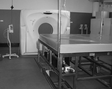
Image Display and Processing
Image Display
CT images map tissue density in the subject. Each pixel in the two-dimensional CT image actually represents a volume of tissue or volume element (voxel) defined by the pixel size in two dimensions and slice thickness in the third dimension. Typical slice thickness ranges from 1 to 10 mm, and now with multislice scanners, images less than 1 mm thick can be obtained. For each voxel, a value is recorded in Hounsfield units (HU) or CT units. This numerical value represents the density of the tissue within the voxel relative to water, which is arbitrarily set to equal zero. Density values of common tissues are listed in Box 20-1. Lesions can be described as hypodense or hyperdense.
BOX 20-1 Density Values of Different Tissues
| Tissue | Density (Hounsfield Units [HU]) |
|---|---|
| Gas | −1000 |
| Fat | −120 |
| Water | 0 |
| Muscle | 40 |
| Equine DDFT | 90-120 |
| Medullary bone | 400-600 |
| Cortical bone | 1500 |
DDFT, Deep digital flexor tendon.
The viewer, using routinely available image review software, can reversibly manipulate the displayed grayscale value of the tissues. Using a wide “window width” is appropriate for the evaluation of tissues with a wide density range such as bone (approximately 400 to 1500 HU). Viewing with a narrow window produces higher image contrast and is appropriate for the evaluation of tissues with a narrow density range such as soft tissue (approximately 40 to 120 HU). “Level” is to the midpoint of the grey shades that are displayed. Changing the level increases or decreases the brightness of the image. In order to perform a complete and thorough evaluation of CT images, the viewer should actively manipulate both window width and level to view all tissues (Figure 20-2).


