Chapter 55 Computed Tomography for the Diagnosis of Sinusitis and Air Sacculitis in Orangutans
Captive orangutans (Pongo pygmaeus, Pongo abelii) have frequently been reported to suffer from upper respiratory tract disease, such as the common cold, sinusitis, and air sacculitis.9,10,13,20 The exact reasons and pathogenesis are unknown until today, and the variety of microorganisms involved is broad. Predisposing factors that are discussed include increased microbacterial contamination of the environment, contact with humans, stress, and long-term antibiotic therapy. In humans, anatomic variations in the nose are the most important predisposing factors for sinusitis, leading to obstruction of the drainage pathways.11,18,23
Although air sacculitis is frequently diagnosed and recognized as a life-threatening disease, sinusitis in orangutans is probably underestimated or even unrecognized because diagnosis is difficult using common diagnostic methods. Also, the clinical signs are often vague. Most evident are chronic nasal discharge, halitosis, cough and, especially in air sacculitis, an enlarged air sac.9,10,13 In recent years, the diagnosis of chronic sinusitis has received more attention, especially because an important correlation to the life-threatening air sacculitis has been suspected.20,24
Because of the inherent uncertainty associated with its diagnosis, more objective tools have been sought. In human medicine, computed tomography (CT) is the most important diagnostic aid to evaluate the upper respiratory tract and follow up on its response to treatment.4,19 CT scanning of the head may represent an advanced imaging technique for the early diagnosis of upper airway diseases, especially of the sinuses, in orangutans.20,24 This would allow the application of treatment modalities to prevent further serious infections of the upper respiratory tract. The increased speed of modern CT techniques and the increased availability of specialized institutions with CT equipment make this important diagnostic imaging technique more readily available to zoological institutions. The purpose of the CT evaluation of the paranasal sinuses and related structures, including the air sacs, is to provide an accurate display of the regional anatomy, confirm the diagnosis, and characterize the extent and localization of disease.
Normal Anatomy and Physiology
The upper respiratory tract in primates includes the nose, larynx, and trachea. In addition to the wide range of size and external shapes of the nose, the internal anatomy and physiology vary considerably between humans and apes, and even between the different ape species.3,14,16,17 A distinct feature of orangutans is the extent of the sinuses and laryngeal air sacs.
The primary function of the orangutan’s nasal airway is breathing and, therefore, in contrast to olfactory-dependent mammals (e.g., carnivores), the anatomy is considerably simpler. A nasal septum in the midline divides the triangular-shaped nasal cavity into two separate passages. Each nasal cavity bears two well-developed scroll-like turbinates, or conchae, the maxilloturbinal inferior (inferior turbinates), and the ethmoturbinal posterosuperior (superior turbinates); the nasoturbinal anterosuperior (middle turbinates) is reduced to a small remnant rostral to the ethmoturbinal.3 The turbinates divide the nasal airway into three groovelike air passages—upper, middle, and lower meatus—and are responsible for forcing inhaled air to flow in a steady regular pattern around the largest possible surface of cilia and climate-controlling tissue. In addition, the nasal passage may be divided into three regions on the basis of their functional significance. The main nasal airway extends from the floor of the nose upward to the middle and superior turbinates and from the tip of the nose backward to the posterior termination of the turbinates. Above the main nasal airway is the olfactory airway. The nasopharynx is the space posterior to the termination of the nasal septum and the turbinates and extends downward to the inferior termination of the soft palate. The nasal cavity is surrounded by two paranasal sinuses (Fig. 55-1), which develop as pneumatized expansions of the nasal cavity’s mucosal recess.8
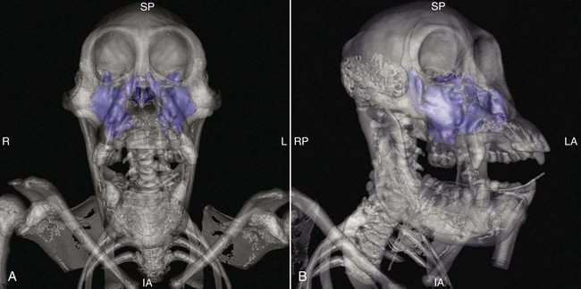
Orangutans differ from other apes in possessing a very narrow interorbital region. The paranasal sinuses include the maxillary and sphenoidal sinuses. Although the maxillary sinus is large, the sphenoidal sinus is significantly reduced in orangutans in contrast to other apes.3 The maxillary sinus occupies the entire maxilla and extends into the lacrimal, frontal, ethmoid, zygomatic, palatine, and sphenoid bones. The sphenoidal extension is situated dorsal to the true sphenoidal sinus and invades both the pterygoid and zygomatic processes of the temporal bone. The nasolacrimal duct, infraorbital, and ethmoidal neurovascular structures pass the maxillary sinus, and the optic nerve and internal carotid artery encroach on its sphenoidal extension.17 The sphenoidal sinuses are found in the most inferior part of the sphenoidal body and may be divided into right and left sinuses separated by an osseous septum. There is no frontal sinus but, in adult orangutans, a frontal recess of the maxillary sinus may extend superiorly to border much of the inferomedial corner of the orbit posterior to the lacrimal duct.8 The extent of this frontal recess is variable, and may sometimes pneumatize the interorbital region of the frontal bone. Before the age of 8 to 12 years, the frontal recess is less developed and the nasal cavity is in direct contact with the medial orbital wall. The maxillary sinuses drain through ostia into the middle meatus, which lies superolaterally to the middle turbinate, whereas the sinus sphenoidalis opens into the upper meatus, which lies lateromedial at the posterior end. All the sinuses are normally in continuous communication with the nasal air.
The nasal cavity and paranasal sinuses are lined with respiratory ciliated epithelium containing serous and mucinous glands.3 The cilia in the respiratory tract are in constant motion and act together to transport the mucus toward the external nasal cavity. In the maxillary sinus, the mucous flow originates along the floor and is centripetally directed toward the ostium, subsequently to the middle meatus, and ultimately to the nasopharynx.5 The flow from the sphenoidal sinus enters the superior meatus and then the nasopharynx.
The larynx is situated in the anterior neck and connects the inferior part of the pharynx with the trachea. It provides a sphincter valve at the entrance to the trachea. The anatomy is similar to that of humans and the laryngeal skeleton consists of nine cartilages—three single (thyroid, cricoid, and epiglottic) and three paired (arytenoid, corniculate, and cuneiform). The hyoid bone is not part of the larynx, although it is connected to it. Slight differences in larynx anatomy have been a key factor in the evolution of human speech.14 A remarkable feature of nonhuman primates is the laryngeal air sac (Fig. 55-2), an accessory mucosal membrane pouch extending from a secondary valve formed by the inferior thyroarytenoid folds.6 Its form and extent vary considerably among apes.15
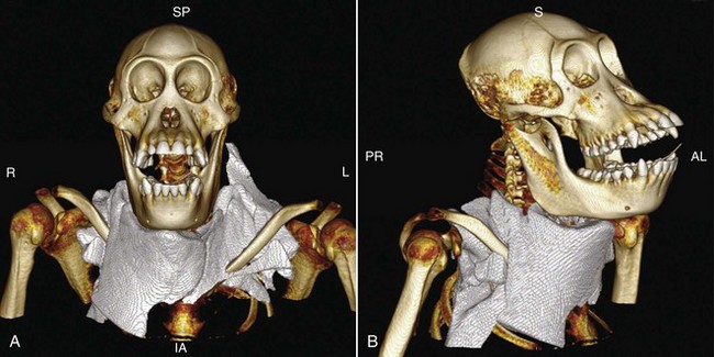
Although there are slight differences in the sac configuration, all the great apes share a rapid expansion of the unilateral sac from the ventricles, which fuse with another sac inferiorly in the late juvenile period.15 The fully developed extralaryngeal air sacs in orangutans extend inferiorly from the ventricles to the neck and axillary region, meet in the midline beneath the pectoral muscles, and are connected to the upper respiratory tract via two openings in the lateral larynx wall. When collapsed, the true capacity of these sacs is difficult to quantify. The air sac is lined with ciliated epithelium with a lamina propria consisting of a layer of loose elastic and collagenous fibers, with variable numbers of seromucinous glands.6 The functions of the laryngeal air sac in primates are still a matter of debate. Suggested functions include storage of expired air to increase oxygen uptake, reduction of the hyperventilation caused by a long sequence of repetitive loud calls, generating another sound source in the laryngeal ventricles, resonating the laryngeal voice source to help produce loud and long calls, and buffering against the pressure induced by intensive expiratory air flow following air trapping during three-dimensional arboreal locomotion. In humans, the saccule is probably the vestigial remains of the air sacs.
Equipment and Computed Tomography Technique
Equipment
CT has been used in veterinary diagnostic imaging since the late 1970s. During a CT scan, a radiographic source rotates around the patient while x-ray detectors are positioned on the opposite side of the circle (gantry) from the x-ray source. Many data scans are progressively taken as the object is gradually passed through the gantry. They are combined together by mathematical procedures known as tomographic reconstruction. Although every slice was formerly taken step by step (sequence CT), newer technologies with faster computer systems and updated calculation software have allowed the production of continuously changing cross sections as the patient to be imaged slowly slides through the gantry within the x-ray circle (spiral CT). Adding multislice detectors to the scanner allows simultaneous scanning of multiple layers at the same time and improved image quality while scan time is reduced (multislice CT). The current newest technology combine two x-ray sources and detectors, which rotate around the patient at the same time and result in better image resolution, higher power, and reduced scan time while the x-ray dose is lowered even further (dual-source CT).21 Reducing the energy dose is important in long-living animals to protect sensitive organs such as the thyroid gland, retina, lens, or gonads from the side effects of the radiation. Repeating a study results in a higher radiation exposure than an adequate energy setup that produces high-quality images.
The images generated are in the axial or transverse plane (slices) and the data of the moving individual slices may be used to generate a three-dimensional picture, viewable from different perspectives. The data obtained represent the varying radiographic intensity sensed at the detectors; through computer calculation, the radiographic density is displayed according to the mean attenuation of the tissue. The attenuation is expressed in Hounsfield units (HU) on a scale from +3071 (most attenuating) to −1024 (least attenuating) HU. Marker points are water, with an attenuation of 0 HU, and air, with −1000 HU; cancellous bone is typically +400 HU and more.21 These Hounsfield units are displayed in terms of relative radiodensity as pixels in a two-dimensional and as voxels in a three-dimensional picture. Contrast medium (e.g., iodine) is sometimes used to highlight structures such as blood vessels to obtain functional information about tissues or to separate tissues of similar attenuation (e.g., mucous membranes and pus).
Stay updated, free articles. Join our Telegram channel

Full access? Get Clinical Tree


