Common Ovine and Caprine Diseases


Common Ovine and Caprine Diseases


Bacterial Diseases


Anthrax


See Chapter 13, Common Bovine Diseases.


Black Disease


Black disease is caused by Clostridium novyi type B. Clostridium novyi is found in soil. Black disease is contracted when its spores are ingested, which commonly occurs while animals are grazing pastures. As the bacteria replicate, they release toxins that cause necrosis of the liver. The exotoxins produced enter the general circulation, where they cause damage to neurons, vascular endothelium, and other vital cells and tissues, leading to sudden death of the animal.


Clinical black disease consists almost entirely of animals found dead. If the animal is identified as being ill before death, it will be depressed, possibly show signs of respiratory distress, and anorexia. Temperature is elevated initially (104–106°F) but declines before death.


Diagnosis of clinical black disease often is made at necropsy. Upon necropsy, hemorrhage of subcutaneous blood vessels is seen, subendocardial and subepicardial hemorrhages are present, and the kidney and liver show signs of autolysis. Migrating flukes, if present, predispose the flock to black disease, and the liver will have channels and show signs of necrosis. Definitive diagnosis can be accomplished with a Gram stain and culture of the liver lesions at the margins of the liver.


Tetracyclines may prove effective if the animal is diagnosed before death, which is rare. Vaccines to prevent black disease are available. Control of liver fluke infestation is extremely important in the prevention of black disease.


Blackleg


See Chapter 13, Common Bovine Diseases.


Botulism


See Chapter 9, Common Equine Diseases.


Brucellosis


Brucellosis in sheep is caused by Brucella ovis. Brucellosis in goats is caused by Brucella melitensis. Brucella melitensis may cause abortion in sheep. Goats can become infected with B. abortus if they are in contact with infected cattle. All of these bacteria are gram-negative coccobacillus organisms. The disease is prevalent in the western portions of North America. It rarely causes abortion in sheep but does cause epididymitis in rams. In sheep the disease is spread from ram to ewe and from ewe to ram, but it cannot be spread from ewe to ewe. Rams can pass the organism in their semen. Goats often become infected once they consume contaminated feed or water. Goats shed the disease in their urine, feces, placenta, and milk.


One of the clinical signs that can be seen in sheep is abortion. If the ewe aborts, she will be free of the disease within a few months. If she does not abort, she will give birth to an infected lamb. The placenta is thickened and necrotic upon examination. Because ewes rarely abort, the major clinical sign is epididymitis in rams. Clinical signs of goats infected with brucellosis include abortion storms, lameness, mastitis, diarrhea, and depression.


Diagnosis is accomplished with the use of agglutination tests or complement fixation.


There is no treatment of brucellosis. Control of the disease usually is accomplished by herd slaughter. To prevent the spread of brucellosis, animals should be serologically tested before they are introduced to the flock. A vaccine is available for sheep, but its ability to prevent the disease is poor Vaccination of goats is not allowed in the United States. Brucella melitensis and B. abortus are considered zoonotic, and precaution should be used when handling potentially infected animals. Only pasteurized milk products should be consumed Brucella melitensis is the cause of Malta fever in humans.


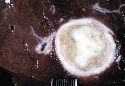
Caseous Lymphadenitis
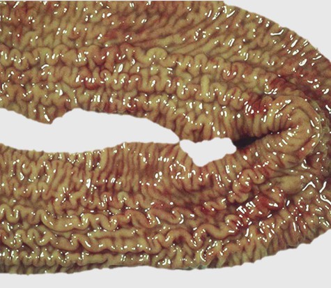

Caseous lymphadenitis is caused by the gram-positive rod Corynebacterium pseudotuberculosis. The bacterium infects both goats and sheep and is often found in manure and soil, on the skin, and in infected organs. The organism can gain access into the body through several different methods, including superficial wounds, ingestion, mucous membranes, and inhalation. Indirect infection can occur through contaminated equipment, including bedding, feeders, and clipper blades.


Clinical signs of infection often include dyspnea, tachypnea, cough, and weight loss.


Diagnosis involves culture of the bacteria from a transtracheal wash. Thoracic radiographs may reveal masses in the thoracic cavity. Hepatic abscesses may be found upon necropsy (Fig. 17-1). The disease often causes abscessation of the lymph nodes. Identification and removal of sick animals from the herd can help control outbreaks. Sanitation during management practices such as castration, tail docking, and parturition can also help prevent infection. Vaccination is controversial.


Chlamydophilosis


Chlamydophilosis is also known as enzootic abortion in ewes (EAE). EAE is caused by the bacterium Chlamydia psittaci. EAE is one of the most common causes of abortion in North America and is the number one cause of abortion in goats. The bacterium is spread by contact with uterine discharge, the fetus, and the placenta. Rams are another source of infection. Ewes infect rams, and the rams then spread the disease to other ewes in the flock. After aborting, the dam develops a good immune response, and elimination of Chlamydia from her uterus usually occurs within 3 months of the abortion, although persistent shedding can occur.


The most common clinical sign of EAE is abortion in the last 2 to 3 weeks of gestation. Lambs can be born weak or stillborn. Chlamydial infections also can produce pneumonia, keratoconjunctivitis, epididymitis, and polyarthritis.


Diagnosis is often achieved through the use of enzyme-linked immunosorbent assay (ELISA) tests, fluorescent antibody staining, and culture isolation.


Antibiotics can be used during the last few weeks of gestation as a form of treatment. Females that have aborted should be segregated from the herd, and any fetal tissue or placenta from the abortion should be burned or buried. Good feeding practices should be followed to help prevent contamination of the feed and water with C. psittaci. Vaccinations are available; however, they most likely will prevent only abortion and not the disease.


Clostridium Perfringens


Clostridium perfringens is a normal flora of the gastrointestinal (GI) system in sheep. The disease (enterotoxemia, overeating disease) can occur in peracute, acute, and chronic forms. All four types of C. perfringens can cause diarrhea in lambs.


Type D


Type D is usually seen in feedlot lambs that consume high-concentrate diets. The disease often affects the lambs with the highest feed-to-gain ratios and in the best condition. It can be seen in animals with abrupt feed changes. The disease may also be seen in lambs that consume excessive amounts of feed or milk. The disease may present with no feeding changes in goats and in vaccinated herds.


Clinical signs include diarrhea, incoordination, excitement, circling, head pressing, convulsions, and sudden death. Sudden death may often occur in sheep without the presence of diarrhea.


Diagnosis before death is often made from clinical signs. Upon necropsy, samples can be taken and cultured from intestinal fluid. The culture can be uninformative because C. perfringens is a normal gut flora and often populates quickly after death. Prevention should include vaccination, control of the parasites, and gradual feed changes. Goats rarely die suddenly without the presence of diarrhea.


Foot Rot


See Chapter 13, Common Bovine Diseases.


Joint Ill


Joint ill primarily occurs in kids. It is caused by several bacterial agents, but the majority are gram-positive agents. Some of the bacteria that cause joint ill include staphylococci, streptococci, Corynebacterium spp., Actinomyces, and coliform bacteria. The bacteria often gain entry to the body through breaks in the skin, the umbilical cord, or the GI or respiratory tract. Predisposing factors include overcrowding and unsanitary conditions.


Clinical signs include warm, painful, swollen joints, lameness, fever, umbilical cord abscessation, and leukocytosis with left shift.


Treatment can be successful if the disease is caught early. Antibiotics and joint flushing can be successful.


Malignant Edema


See Chapter 13, Common Bovine Diseases.


Pinkeye


See Chapter 13, Common Bovine Diseases.


Transmissible Spongiform Encephalopathies


Transmissible spongiform encephalopathies (TSE) is also known as “scrapie.” Scrapie is thought to be caused by a prion. Sheep seem to be more or less sensitive to TSE depending on their genetics. Suffolk sheep are most commonly diagnosed. Sheep and goats are natural reservoirs for scrapie. Transmission of the disease is not fully understood. Sheep are suspected to become infected at birth and often begin displaying clinical signs around image years of age. The infection is thought to be result from contact with infected animals or the environment.


Clinical signs include wool or hair loss, ataxia, weight loss, starring, aggressiveness, floppy ears, tremors, seizures, inability to swallow, and death. The disease is chronic and degenerative.


Diagnosis is accomplished by finding the scrapie prion protein with immunostaining of lymphoid tissue. There is no treatment of scrapie. The disease is considered fatal. The disease is reportable and often results in flock slaughter.


Prevention is being attempted through the Scrapie Flock Certification Program. The program requires flocks to be assessed for 5 years to determine that they do not contain any scrapie-infected sheep. This program has four levels of clearance. When the final level is attained, the producers are able to export and sell sheep both within the United States and internationally without restrictions.


Other Microbial Diseases
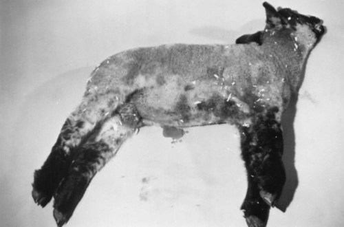

Toxoplasma


Toxoplasma gondii is a protozoan that causes abortion in sheep and goats but only rarely causes abortion in cattle and horses. Infection with toxoplasma results in fetal death and abortion, embryonic death, stillbirth, or birth of weak, nonviable lambs or kids. Placental infection occurs approximately 14 days after ingestion of oocysts. Most abortions take place 1 month before parturition.


The most common evidence of toxoplasma is white, chalky foci of necrosis and calcification up to 2 mm in diameter in cotyledons. Tachyzoites may be found in placenta or other fetal tissues but are not numerous. Several serologic tests, including the modified agglutination test, indirect fluorescent antibody test, Sabin-Feldman dye test, indirect hemagglutination test, and ELISA, reliably detect toxoplasmosis in pleural or amniotic fluid or presuckling serum from nondecomposed fetuses. Aborted tissues may be infectious to humans and should be handled with caution.


Ewes or does are often infected by age 4 years. They seldom abort from toxoplasmosis in subsequent pregnancies. Prevention should include reducing cat access to sheep areas and equipment as well as preventing cats from eating placenta and tissue (Tables 17-1 and 17-2).


TABLE 17-1


Sheep Vaccinations*


Disease/Vaccination Ewes Lambs Feedlot lambs Rams Comments
Clostridium perfringens type C 4–6 weeks before parturition
If animals have never been vaccinated, twice 4 weeks apart with last dose 4–6 weeks before parturition
If born to unvaccinated ewe, at birth and booster in 4–6 weeks
Lambs from vaccinated ewes should be vaccinated at 12–16 weeks and booster given in 4–6 weeks
Upon entering feedlot and booster in 2–4 weeks Annually  
Clostridium perfringens type D 4–6 weeks before parturition
If animals have never been vaccinated, twice 4 weeks apart with last dose 4–6 weeks before parturition
If born to unvaccinated ewe, at birth and booster in 4–6 weeks
Lambs from vaccinated ewes should be vaccinated at 12–16 weeks and booster given in 4–6 weeks
Upon entering feedlot and booster in 2–4 weeks Annually  
Clostridium tetani Can be given during pregnancy with Clostridium types C and D At time of castration and tail docking Annually Annually Often combined with Clostridium types C and D
Other clostridial diseases (black disease, blackleg, malignant edema, struck, lamb dysentery, botulism) 4–6 weeks before parturition
If animals have never been vaccinated, twice 4 weeks apart with last dose 4–6 weeks before parturition
If born to unvaccinated ewe, at birth and booster in 4–6 weeks
Lambs from vaccinated ewes should be vaccinated at 12–16 weeks and booster given in 4–6 weeks
Annually Annually Primarily used only in high-risk herds
Leptospirosis         Primarily used only in high-risk herds
Sore mouth At least 2 months before parturition, booster every 5–12 months depending on risk 1–2 days of age, booster every 5–12 months depending on risk 4 weeks before risk, booster every 5–12 months depending on risk 4 weeks before risk, booster every 5–12 months depending on risk Live virus
Vaccinated sheep can spread the disease for up to 8 weeks after vaccination
Use in infected herds only
Performed by scratching skin in area without wool (inner ear or under tail in adults and inner thigh in young animals) and then brushing on the vaccine
Sores will form at application site
Foot rot 4 weeks before lambing, booster every 4–6 months 4 weeks of age, booster in 4–8 weeks 4 weeks before wet/rainy season, booster every 4–6 months 4 weeks before wet/rainy season, booster every 4–6 months Vaccinate behind the ear
Only reduces infection levels
Abscesses are not uncommon, discoloration of the wool at the injection site
Booster in 4 weeks from first time of vaccination
Caseous lymphadenitis Annually Annually Annually Annually Primarily used only in high-risk or infected herds
Booster in 4 weeks after the first dose
Enzootic abortion in ewes (EAE) 4 weeks before breeding
Do not use in pregnant ewes
      Primarily used only in high-risk or infected herds
Toxoplasma 4 weeks before breeding
Do not use in pregnant ewes
Booster every 2 years
      Primarily in only high-risk or infected herds.
Vaccine is not available in the United States
Vibriosis Annually, 2 weeks before breeding
Booster in midpregnancy if first vaccination
      Primarily used only in high-risk herds
Brucellosis       Rams test positive if vaccinated  
Rabies         Common in pet sheep and possibly in endemic areas
Escherichia coli 4–6 weeks before parturition
If animals have never been vaccinated, twice 4 weeks apart
If ewes were unvaccinated, oral antibody can be given at birth     Primarily used only if diarrhea in 1- to 2-day-old lambs is a problem

Only gold members can continue reading. Log In or Register to continue

Stay updated, free articles. Join our Telegram channel

Aug 11, 2016 | Posted by in INTERNAL MEDICINE | Comments Off on Common Ovine and Caprine Diseases

Full access? Get Clinical Tree

Get Clinical Tree app for offline access