Colorectal neoplasia in a dog

38 Colorectal neoplasia in a dog


Physical examination


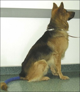
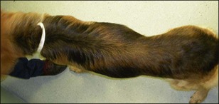
The dog was bright, alert and responsive (Fig 38.1) although he was quite thin (body condition score 3/9) with muscle loss over the lumbar area (Fig 38.2). His mucous membranes were pink and moist with a capillary refill time of <2 seconds and hydration appeared adequate. No abnormalities were noted on thoracic auscultation. His heart rate was 82 beats per minute with good matched pulses and respiratory rate was 32 breaths per minute. Peripheral lymph nodes were normal in size. Due to the history of haematochezia, taking the rectal temperature was delayed until analgesia (e.g. topical lignocaine) could be used. When taken later it was 38.2° C.


On abdominal palpation several large firm masses were identified. One of these was especially prominent in the caudal abdomen and was felt to be at least 4 to 5 cm in diameter. The masses were not attached to the body wall and could be moved fairly freely. A tubular mass was present in the ventral abdomen, which was thought to be either a ventrally displaced colon or an enlarged loop of small intestine. The dog appeared uncomfortable during abdominal palpation.


Problem list and discussion of problems


The dog’s primary problems were haematochezia and tenesmus consistent with large intestinal disease. The frequency and urgency of the diarrhoea was also consistent with disease of the colon and/or rectum, although weight loss is less common in large intestinal disease.


Severe acute pancreatitis can cause melaena and haematochezia, but this dog was too bright to have severe acute pancreatitis. The dog had been given an adequate dose of fenbendazole, making gastrointestinal parasites less likely. While he had previously been on meloxicam, there had been a period of about a month where it had not been given and there had been no change in clinical signs.


The other problem was the presence of masses or thickened areas found during palpation of the abdomen. The most likely causes of these were thought to be neoplasia, lymph node enlargement or intussusception(s).

Stay updated, free articles. Join our Telegram channel

Sep 22, 2016 | Posted by in SMALL ANIMAL | Comments Off on Colorectal neoplasia in a dog

Full access? Get Clinical Tree

Get Clinical Tree app for offline access