20 Clinical lesions and signs of discomfort
ORAL EXAMINATION – UNDER GENERAL ANAESTHESIA
In summary, examination under general anaesthesia identified the following:
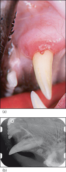
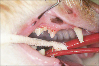
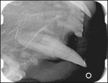
3. Small cavities at the buccal gingival margin of 204 (Fig. 20.3a) and 107 (Fig. 20.4). The lesion on 107 was only obvious after calculus had been removed.
4. Teeth 106, 206, 207 and 407 were missing. The gingiva at these sites was not inflamed except over 407, which was intensely inflamed.
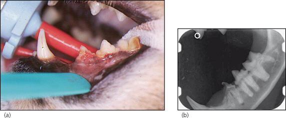
Figure 20.1 Lateral photograph (a) and lateral radiograph (b) of the left lower quadrant.
(b) The radiograph shows that external root resorption has caused destruction and bone replacement of the mesial root. In contrast, the distal root is radiographically unaffected. The resorptive process has spread to involve the crown, resulting in the obvious clinical lesion. Once the disease is clinically apparent, i.e. a cavity is formed, root resorption is extensive. The first clinically detectable sign of resorption is already an end-stage lesion.
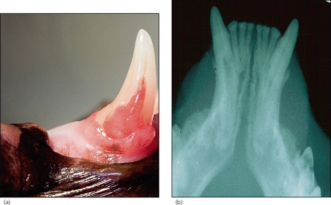
Figure 20.2 Lateral photograph of the right lower canine (a) and radiograph of the rostral lower jaw (b).
