13 Chronic gingivostomatitis associated with FeLV and FIV
ORAL EXAMINATION – UNDER GENERAL ANAESTHETIC
In summary, examination under general anaesthesia identified the following:
1. Teeth 204, 208, 209, 107, 108, 109, 309 and 409 were missing (presumably extracted 10 weeks earlier)
4. Gingivostomatitis of alveolar and buccal mucosa bilaterally in the edentulous upper premolar and molar regions (Fig. 13.3).
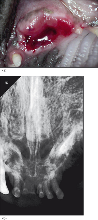
Figure 13.1 Occlusal photograph (a) and rostrocaudal radiograph (b) of the non-healing extraction socket of 204.
(a) The socket has not healed after extraction of 204 10 weeks earlier. In fact, the owner reported that the defect was larger than it was immediately after the extraction.
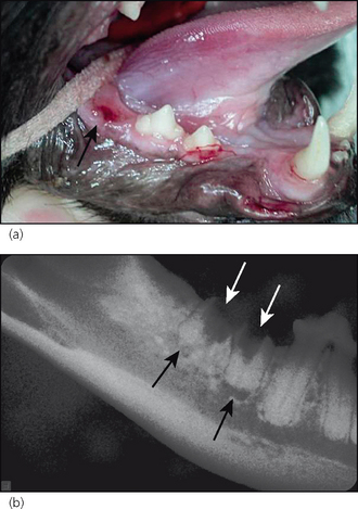
Figure 13.2 Lateral photograph (a) and radiograph (b) of the right mandibular quadrant.
Stay updated, free articles. Join our Telegram channel

Full access? Get Clinical Tree


