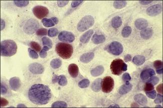
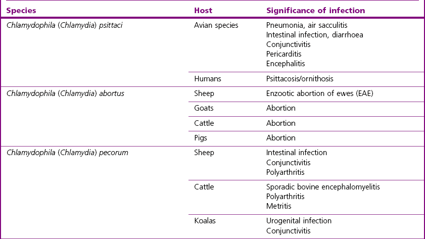
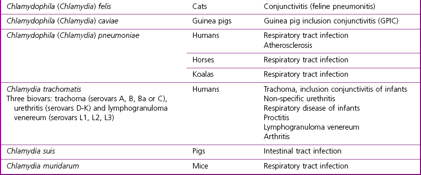
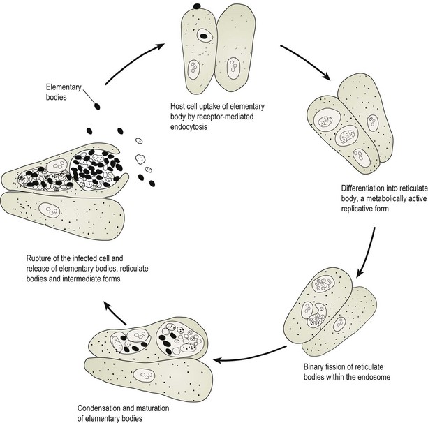
Chapter 33 This large group of obligate intracellular bacteria is characterized by a unique, biphasic developmental cycle involving replication in cytoplasmic inclusion vacuoles within eukaryotic cells (Fig. 33.1). The order Chlamydiales consists of four families, Chlamydiaceae, Parachlamydiaceae, Simkaniaceae and Waddliaceae. A rapid expansion in the number of organisms within the order has occurred in recent years to include members from numerous animal species including invertebrates and environmental sources. Taxonomic changes have accompanied the expansion of the order (Everett et al. 1999). Neochlamydia hartmannellae, an amoebic endosymbiont, has been associated with ocular disease in cats (von Bomhard et al. 2003). Waddlia chondrophila and Parachlamydia species have been implicated in bovine abortion (Ruhl et al. 2009). From a veterinary and medical point of view, the most significant species are found in the family Chlamydiaceae. Currently nine species are recognized. There is on-going debate regarding the allocation of these species to a single genus (Stephens et al. 2009, Greub 2010a, 2010b, Kuo et al. 2011) or to two genera (Everett & Andersen 1999, Everett et al. 1999). Phylogenetic analyses (Voigt et al. 2012) support the recognition of Chlamydophila species as a distinct evolutionary branch (Table 33.1). Chlamydiae have been termed ‘energy parasites’ on account of their apparent inability to generate ATP and resulting dependence on the host cell to supply NTPs. Both plasmids and bacteriophages are found in chlamydiae. The developmental cycle of chlamydiae involves morphologically distinct infectious and reproductive forms (Figs. 33.1 and 33.2). The infectious extracellular form, termed the elementary body (EB), is small (200–300 nm), metabolically inert and osmotically stable. The EB is surrounded by a conventional bacterial cytoplasmic membrane, a periplasmic space and a lipopolysaccharide-containing outer envelope. The periplasmic space does not contain a detectable peptidoglycan layer and the EB relies on disulphide cross-linked envelope proteins for osmotic stability (Hatch 1996). The EB enters a host cell by receptor-mediated endocytosis. Acidification of the endosome and subsequent phagolysosomal fusion is prevented by a mechanism that is not fully understood. A process of reorganization lasting several hours follows and results in the conversion of the EB into the reticulate body (RB). The RB is about 1 µm in diameter, metabolically active, osmotically fragile and divides by binary fission to produce more RBs. In cells infected with C. trachomatis and containing more than one inclusion, fusion of the inclusions is possible. At about 20 hours post infection the cycle becomes asynchronous with some RBs continuing to divide while others condense and mature to form EBs. In general the cycle continues till about 30–72 hours after infection when the host cell lyses, releasing several hundred EBs, RBs and intermediate forms. Under stressful conditions such as nutrient deprivation, particularly shortages of the amino acids tryptophan and cysteine, certain immunological responses such as the presence of gamma interferon or exposure to antibiotics such as penicillin, chlamydial replication may become suspended resulting in a state of persistent infection and the appearance of large, aberrant RBs. Delayed infections of this nature appear to be important in the development of immunopathology, associated with diseases such as trachoma and pelvic inflammatory disease in humans. Figure 33.2 Immunoperoxidase staining with haematoxylin counter stain. Chlamydophila (Chlamydia) abortus (enzootic abortion of ewes) intracellular inclusions (red-brown) in McCoy cells. (×400) Many chlamydial infections are asymptomatic, particularly where the infections are localized to superficial epithelial or mucosal surfaces, and may persist for long periods while inducing little protective immunity. Chronic, recurrent infections may provide an opportunity for repeated stimulation of the host immune system with particular chlamydial antigens. In a number of diseases such as trachoma or pelvic inflammatory disease in humans the pathology encountered is more severe than would be expected from infection alone. Chlamydiae possess a number of heat shock proteins which are similar to heat shock proteins in other bacteria and to a number of human mitochondrial proteins. It is believed that chronic stimulation of the immune system with these proteins contributes significantly to the delayed type hypersensitivity responses associated with tubal or conjunctival scarring in pelvic inflammatory disease and trachoma, respectively. Gamma interferon is produced in response to chlamydial infection and has been shown to be chlamydiastatic. It is likely that gamma interferon is important in helping to control primary chlamydial infections. However, there is also evidence that gamma interferon is responsible for the induction of a state of latency or persistence in chlamydial infections which in turn may be responsible for up-regulation of heat shock protein expression (Ward 1995). A very wide range of species of animals, including invertebrates, are known to be susceptible to infection with chlamydiae (Vanrompay et al. 1995). In mammals and birds, both the severity and the type of disease produced by chlamydiae are highly variable, ranging from clinically inapparent infections to local infections of epithelial surfaces to systemic infections spread by the bloodstream (Table 33.1). In mammalian species chlamydiae have been associated with intestinal infection, gastritis, pneumonia, abortion, urogenital infection, mastitis, polyarthritis, polyserositis, conjunctivitis, encephalomyelitis and hepatitis. In avian species chlamydiae are associated with generalized infection, pneumonia, air sacculitis, pericarditis, encephalitis, diarrhoea and conjunctivitis. A single clinical presentation usually predominates in an outbreak or series of related clinical cases. The factors influencing the clinical signs and their severity include: • Species or strain of chlamydia. In ruminants, C. abortus has a tropism for the trophoblast cells of the placenta and is an important cause of abortion, whereas infections with C. pecorum are frequently inapparent. There are at least six avian serotypes of C. psittaci labelled A through F. The syndromes and host range of particular serotypes are fairly specific: serotype A, psittacine birds; serotype B, pigeons; serotype C, ducks; serotype D, turkeys; serotype E, pigeons and ratites; and one isolate of serotype F from a psittacine bird (Andersen 1991, 1997). The serotypes can be distinguished using serovar-specific monoclonal antibodies or the PCR-RFLP technique (Vanrompay et al. 1997). Three further genotypes are generally accepted: genotype E/B, group of isolates from ducks, turkeys and pigeons; and two mammalian isolates, WC in cattle and M56 in muskrats (Geens et al. 2005). Based on sequence analysis of the ompA gene a further six genotypes of C. psittaci have been described (Sachse et al. 2008). • Species of host. Chlamydial strains are associated with specific diseases in particular hosts. Interspecies transmission is uncommon. Disease in a secondary host may be similar as occurs when C. abortus is transmitted from sheep to catttle or it may be more severe as can occur when C. abortus transmits from sheep to pregnant women. • Age, sex and physiological state. Lambs usually acquire C. abortus infections early in life on endemically infected farms. However, clinical signs do not become apparent until the animal is pregnant. Clinical signs are rare in infected rams. • Route of infection and degree of exposure to chlamydiae. Chlamydophila (Chlamydia) pecorum is associated with conjunctivitis, arthritis and inapparent intestinal infection in sheep. • Environment and management practices. Enzootic abortion of ewes is a disease of lowland, intensively managed flocks where indoor lambing facilitates the spread of infection to neonates and in-contact ewes. Chlamydophila (Chlamydia) psittaci strains are transmissible to man with varying efficiency. Isolates of C. psittaci are considered the most zoonotic of the chlamydial pathogens of animals but human infections may also be acquired following contact with aborting ewes (C. abortus) or cats with conjunctivitis (C. felis). Human infections acquired from psittacine species are termed psittacosis while those acquired from other avian species are termed ornithosis. There is no significant difference in the clinical course of either condition. Infection is usually acquired following inhalation of aerosols with infections varying from inapparent to severe systemic disease. Pulmonary involvement is common. Infected humans typically have a cough, headaches, fever and myalgia. Severe cases may develop meningitis or meningoencephalitis. Death is rare provided adequate treatment is received. Diagnosis is usually based on the collection of paired serum samples and the demonstration of a significant rise in antibody titre by CFT or immunofluorescence. Psittacosis is a notifiable disease in many European countries as well as the USA and Australia. Chlamydophila abortus is a serious and potentially life-threatening zoonosis in pregnant women (Johnson et al. 1985, Buxton 1986). Pregnant women should avoid all contact with lambing ewes.
Chlamydiales

Pathogenesis and Pathogenicity
Clinical Infections
![]()
Stay updated, free articles. Join our Telegram channel

Full access? Get Clinical Tree


