Anthony P. Pease
Cerebrospinal Fluid Standing Tap
Cerebrospinal fluid (CSF) collection is a valuable aid for reaching a diagnosis in the horse with neurologic signs, such as those in which equine protozoal myeloencephalitis is suspected. Two main sites are available for collection of CSF: the lumbosacral cistern and the cerebellomedullary cistern in the cervical region. The lumbosacral procedure is usually performed under local anesthesia with the horse standing, but cervical centesis of the cerebellomedullary cistern through the atlanto-occipital space necessitates general anesthesia, which can put the horse with neurologic dysfunction at risk for injury during recovery. For the latter reason, the lumbosacral approach is usually used to obtain CSF samples from horses with neurologic disease, even if a sample from the cerebellomedullary cistern might be more diagnostic. Recently, a new approach to the cerebellomedullary cistern has been described for use in standing horses. The procedure, which involves making a lateral approach between C1 and C2, offers a convenient and safe alternative to the lumbosacral procedure in the standing horse.
Lumbosacral Procedure
The lumbosacral cistern is a focal accumulation of CSF in the caudal part of the vertebral canal. The site is approached with an 18-gauge, 15.2-cm (6-inch) needle introduced just cranial to a divot that can be palpated cranial to the tuber sacrale between L6 and S1. In a large horse, such as a warmblood, a 20-cm (8-inch) needle may be needed. Identification of the junction between L6 and S1 in both the sagittal and transverse planes can be assisted by ultrasonography, with a 2- to 5-MHz macroconvex transducer imaging approximately 12 cm in depth. A 7.5-MHz microconvex probe can also be used, but the image quality is reduced because of the depth required to image the level of the vertebral canal. The horse is generally placed in stocks and sedated with detomidine hydrochloride (0.01 mg/kg, IV) and butorphanol tartrate (0.01 mg/kg, IV). Iatrogenic blood contamination of CSF from a lumbosacral puncture is common, especially in horses with neurologic signs. The source of this blood could be from damage to the epidural vein, meningeal vessels, or spinal cord vessels, or the large mass of muscle tissue that is traversed to obtain the sample. Applying gentle suction and discarding the first few milliliters of CSF will aid in obtaining an appropriate sample because iatrogenic hemorrhage usually clears quickly. Occasionally the needle needs to be replaced or the procedure has to be repeated at a later time to allow the hemorrhage to cease. The other possible complication is a potentially violent reaction that can occur during needle puncture. Because the tip of the needle cannot be observed during the procedure, it is possible that puncture of a nerve root or traversing a portion of spinal cord causes this reaction. When this occurs, the needle must be removed and the procedure repeated either immediately or at a later time.
Cervical Procedure
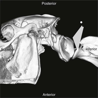
Standing cervical centesis obtained dorsally through the atlanto-occipital region into the cerebellomedullary cistern was first described in 1968. The chief problem with this approach is the high risk for a horse raising its head and causing the needle to enter the spinal cord. In one instance, a horse collapsed during the procedure, but regained consciousness in about 2 hours. A safer approach is to use ultrasound guidance and obtain cerebrospinal fluid from the C1-C2 articulation. Examination of the skeleton of the horse shows that dorsal to the dens and ventral to the cranial aspect of the C2 spinous process is a 3-cm region of spinal cord that is exposed (Figure 84-1). A lateral approach with a 4- to 10-MHz microconvex or a 7.5- to 12-MHz linear probe allows for easy evaluation of the spinal cord at this site. Because the spinal cord is only 6 cm below the skin, an 18-gauge, 8.9-cm (3.5-inch) spinal needle can be used to obtain a sample of CSF. The needle size and procedure are independent of the size of the horse, and the procedure has been performed on a draft horse weighing 773 kg (1700 pounds). Horses can be placed in a stock, or the procedure can be performed stallside; it has even been performed on horses that are laterally recumbent and unable to stand. At the time of this writing, the procedure had been performed without immediate complications in nearly 20 horses with neurologic signs.

< div class='tao-gold-member'>
Stay updated, free articles. Join our Telegram channel

Full access? Get Clinical Tree


