Chapter 143 Cardiovascular Radiography
Thoracic radiography and echocardiography are useful in the recognition and assessment of cardiovascular diseases. Other imaging modalities, such as cardiac magnetic resonance imaging, gated computerized tomography, and radionuclide studies, are uncommonly used in routine cardiology practice and have limited indications at this time. This chapter emphasizes the use of radiography in cardiac diagnosis.*
When combined with the results of the clinical examination, thoracic radiographs may contribute to the specific cardiac diagnosis, verify the presence of congestive heart failure (CHF), and assist in the differential diagnosis of respiratory signs such as coughing and dyspnea. Analysis of thoracic radiographs in the diagnosis of respiratory diseases is discussed in more detail in Chapter 159. Essential principles in the application of echocardiography are considered at the end of this chapter. More specific applications of radiography and echocardiography are discussed in other chapters throughout this section.
GENERAL PRINCIPLES OF CARDIAC RADIOGRAPHY
Technical Considerations
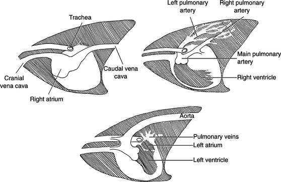
Normal Lateral Radiograph (Fig. 143-1)
Dogs
The following are guidelines for understanding the normal canine lateral thoracic radiograph:
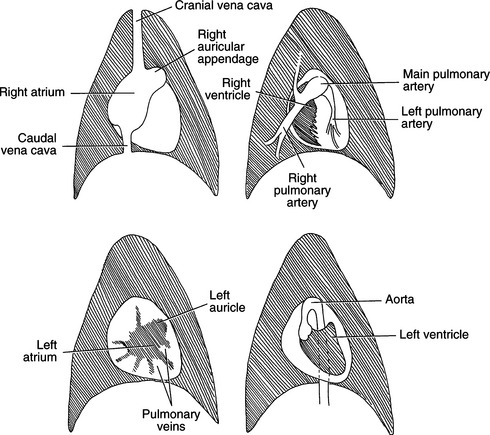
Normal Ventrodorsal or Dorsoventral Radiograph (Fig. 143-2)
Dogs
The following are guidelines for understanding the normal canine VD or DV thoracic radiograph:
Normal Pulmonary Arteries and Veins
Location and Size
INTERPRETING THORACIC RADIOGRAPHS IN CARDIAC DISEASE
Once an adequately positioned and exposed radiograph has been obtained, the radiographs must be interpreted in a systematic manner. Optimally, the films would be assessed without knowledge of the patient and then reassessed after reading the signalment, clinical history, and examination findings. In most instances this is impractical, but it is clear that including the clinical findings will both assist the evaluation (by providing a clinical perspective) and hinder an objective assessment (by prejudicing the reader with the clinical diagnosis).


