Chapter 20 Cardiovascular Disease, Lymphoproliferative Disorders, and Thymomas
Part I Cardiovascular Diseases
Normal Cardiovascular Structure
The rabbit heart is unique in several ways: the tricuspid valve is composed of two rather than three cusps; the aortic nerve is not associated with chemoreceptors but only with baroreceptors; the rabbit pulmonary artery and its branches are heavily muscular6; and a persistent left cranial vena cava is also normally found and drains into the coronary sinus.25 Additionally, the myocardium has limited collateral circulation and is therefore predisposed to ischemia mediated by coronary vasoconstriction.36
Examination of the Rabbit with Cardiovascular Disease
History
A thorough general history, including husbandry, diet, and past or present illnesses, should be obtained for rabbits suspected of having cardiac disease. Tachypnea, dyspnea, syncope, anorexia, weight loss, and malaise may be signs of heart disease in rabbits.
Physical Examination
Take care to minimize the handling of a rabbit in cardiac distress; a complete physical examination and further diagnostic testing must sometimes be delayed until the rabbit is clinically stable. Stress can be minimized by examining the rabbit in a quiet room and moving slowly. Rabbits are a prey species and thereby very sensitive to external stimuli. If necessary, sedatives may also be given to minimize the risk of self-injury. Sedation can be achieved with midazolam 0.5 to 1 mg/kg intramuscularly45 or, for heavier sedation, a combination of ketamine 5 to 20 mg/kg and midazolam 1 to 2 mg/kg subcutaneously13 with minimal cardiovascular depression (see Chapters 31 and Appendix). Provide flow-by oxygen as needed if a rabbit is dyspneic or becomes tachypneic with handling. In rabbits suspected of having cardiac disease or respiratory distress, focus the initial examination on observing the respiratory rate and pattern, obtaining the heart rate and rhythm, auscultating the thorax, examining the mucous membranes, and palpating the pulses. Normal heart rate is 180 to 250 beats per minute and normal respiratory rate is 30 to 60 breaths per minute. Peripheral pulses can be palpated at the central artery of the ear and should be strong and synchronous with the heartbeat. Two heart sounds (S1 and S2) are normally heard. Rabbits are obligate nose breathers; therefore do not occlude the nares.
Diagnostic Methods
Radiography
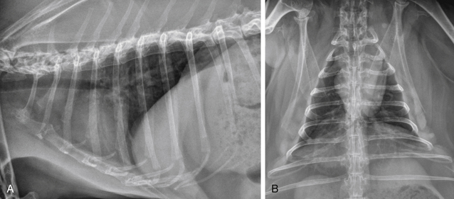
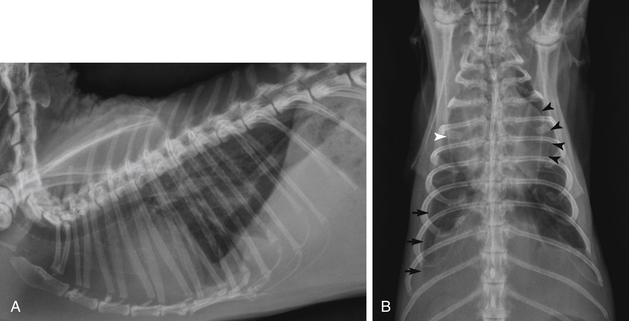
Thoracic radiography provides critical information in the patient with cardiopulmonary disease—namely cardiac shape and size, pulmonary pattern, vascular pattern, and other thoracic lesions. Congestive heart failure and respiratory disease can be differentiated by evaluating thoracic radiographs. Radiographic findings supporting a diagnosis of cardiac disease are similar to those in other species and include cardiac enlargement, pulmonary vascular enlargement, pulmonary interstitial and alveolar pulmonary pattern of pulmonary edema, and pleural effusion. Figures 20-1 and 20-2 show thoracic radiographs of a normal adult rabbit and a rabbit with heart disease, respectively.
Electrocardiography
Electrocardiography (ECG) is a simple and practical diagnostic test in rabbits with suspected or confirmed cardiac disease. An ECG is critical to diagnose and manage arrhythmias or syncope. The ECG may also be a helpful addition to the cardiac database. However, one should not use an ECG to assess or detect chamber enlargement or hypertrophy. Record the ECG with the rabbit in sternal recumbency. File the alligator-style ECG clips to minimize skin trauma, and place medical-grade alcohol or electrode gel on the clips to enhance conduction. Record tracings with a vertical calibration of 2 cm/mV and a horizontal paper speed of 50 mm/s. Normal rabbit rhythm is sinus and does not include respiratory sinus arrhythmia.44 Spontaneous changes in the QRS complex have been observed in normal rabbits in serial ECG recordings. Normal ECG values for a variety of pet rabbit breeds have been reported.34,45 In the authors’ experience, R-wave amplitudes in lead II of 0.1 to 0.2 mV seem most common. Reference ranges for ECGs are summarized in Table 20-1.
Table 20-1 Electrocardiographic Values in Clinically Normal Pet Rabbits
| ECG Parameter | Values34 | Values45 |
|---|---|---|
| Heart rate | 198-330 (mean 264) beats/minute | 240 beats/minute (mean) |
| Measurements (lead II) | ||
| P wave | ||
| Amplitude (height) | 0.04-0.12 mV | 0.04-0.07 mV |
| Duration (width) | 0.01-0.05 s | 0.02-0.04 s |
| P-R interval | ||
| Duration | 0.04-0.08 s | 0.05-0.07 s |
| QRS complex | ||
| R-wave amplitude | 0.03-0.039 mV | 0.12-0.2 mV |
| Duration | 0.02-0.06 s | 0.03-0.04 s |
| Q-T interval | ||
| Duration | 0.08-0.16 s | — |
| T wave | ||
| Amplitude | 0.05-0.17 mV | — |
| Electrical axis (frontal plane) | 43-80 degrees | — |
| Body weight | 1.1-7.9 (mean 2.57) kg | — |
Echocardiography
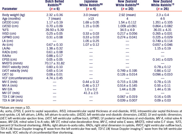
Echocardiography provides a sensitive, accurate, noninvasive means of assessing the heart. Most rabbits tolerate echocardiography easily, making it a practical diagnostic tool. However, in rabbits that are tachypneic or dyspneic, give oxygen by face mask during restraint. The echocardiographic exam may be performed in lateral or sternal recumbency. Standard views used to evaluate dogs and cats can also be obtained with the rabbit. Because of the rabbit’s rapid heart rate and small size, optimal evaluation requires a high-frequency transducer and high-frame-rate ultrasound machine. Two-dimensional and M-mode echocardiography assess cardiac structure, chamber size, wall thickness, and motion as well as extracardiac structures, masses, and pleural effusion. Color-flow, spectral, and tissue Doppler echocardiography assess direction and velocity of blood flow, further defining cardiac conditions with more insight on systolic and diastolic function. Normal echocardiographic values have been published for several breeds of rabbits (Table 20-2). Sedative drugs used for restraint may affect cardiac measurements, especially alpha2-agonists such as dexmedetomidine and xylazine. Reduced percent fractional shortening, E/A reversal, decreased heart rate, and other changes have been documented with intramuscular ketamine at dose of 50 mg/kg and xylazine at 4 mg/kg.54 Sedation with ketamine 20 mg/kg and midazolam 2 mg/kg given subcutaneously has been shown to be less cardiodepressive.13
Diseases and Management
Congestive Heart Failure
Long-term therapy of CHF should include a diuretic (furosemide 1-2 mg/kg PO q8-24h) combined with treatment directed at the underlying precipitating cause. Knowledge of the cardiac disease process is the basis of specific treatment of the underlying condition. No cardiac drugs are approved for use in rabbits by the U.S. Food and Drug Administration. Drug dosages for rabbits are not available for all cardiac medications, but drugs and dosages published for cats or ferrets may be successfully used on a milligram-per-kilogram basis. Angiotensin-converting enzyme inhibitors such as enalapril maleate (0.25-0.5 mg/kg PO q24-48h, begun q24h) may be beneficial in treating rabbits with congestive heart failure. Pimobendan (0.1-0.3 mg/kg PO q12-24h) may also be used for treatment of systolic dysfunction and myocardial failure.45 During acute and chronic management, it is critical that clinical and radiographic signs, hydration status, appetite, and body weight as well as serum or plasma blood urea nitrogen, creatinine, and electrolyte concentrations be monitored. For drugs and dosages, refer to Chapter 41.
Congenital Heart Disease
Congenital heart disease in rabbits is rarely reported. Ventricular septal defect, diagnosed with echocardiography, has been described.47 A ventricular septal defect, pulmonary hypertension, and valvular cyst identified at necropsy have been described in a New Zealand white rabbit.31
Arrhythmia
Arrhythmias such as atrial fibrillation and ventricular premature complexes have been identified in pet rabbits with underlying cardiomyopathies and congestive heart failure.35 We have also diagnosed arrhythmias in several rabbits that exhibited syncope and an irregular heartbeat. In a rabbit with an arrhythmia, the treatment protocol should be based on ECG findings and clinical signs. For syncope associated with bradycardia, oral theophylline or pacemaker implantation may be indicated. Glycopyrrolate may be more effective than atropine sulfate in increasing heart rate.43 Atropine may not be as effective because some rabbits produce atropinesterases. Supraventricular tachycardias may be treated with oral digoxin or diltiazem. Ventricular tachyarrhythmias may respond to intravenous lidocaine. Dosages are published for lidocaine, verapamil, atropine, and glycopyrrolate. Other antiarrhythmic drugs may be used at dosages published for cats or ferrets.
Myocardial Disease
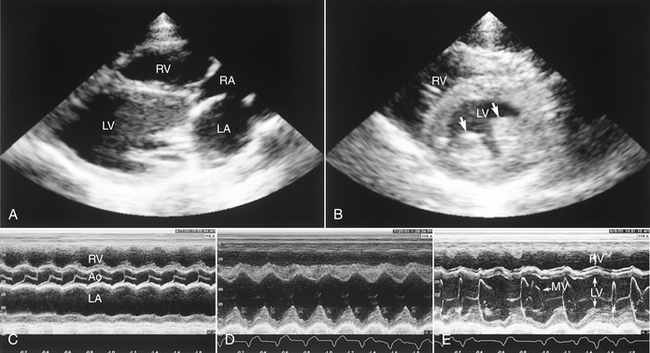
Numerous myocardial diseases have been reported in rabbits, and cardiomyopathy is a common postmortem finding in older rabbits.47 Idiopathic hypertrophic cardiomyopathy and dilated cardiomyopathy have been diagnosed by echocardiography (Fig. 20-3).44 Vitamin E deficiency produces a muscular dystrophy in which the myocardium may be affected.3 In experimental studies, myocardial disease has been created through inoculation of Trypanosoma cruzi49 and administration of doxorubicin.59 Infectious myocardial diseases are rare in pet rabbits. Known infectious organisms include Pasteurella multocida, Salmonella species, Encephalitozoon cuniculi,36 and coronavirus.8 The alpha-agonist drug detomidine has been associated with myocardial necrosis and fibrosis in New Zealand white rabbits.23 A similar ischemia-mediated process is suggested in association with ketamine/xylazine administration.36
Valvular Disease
Mitral and tricuspid insufficiencies are not uncommon and have been identified in pet rabbits.44 Valvular insufficiencies may be associated with primary valve degeneration, cardiomyopathy, or infection. Progression of the condition leads to volume overload and potential congestive heart failure. A focal murmur is the most common clinical finding. Valvular disease is diagnosed with two-dimensional and Doppler echocardiography (see Fig. 20-3). Echocardiographic findings most often include thickening of one or both atrioventricular valves, dilation of cardiac chambers, and turbulent regurgitation of blood detected by Doppler.
Valvular endocarditis, caused by Staphylococcus aureus, has been reported in rabbits.53
Vascular Disease
Spontaneous arteriosclerosis of the aorta and other arteries has been observed in nearly all rabbit breeds. Clinical signs, if present, may include lethargy, anorexia, and weight loss. The cause is unknown. In rabbits with spontaneous arteriosclerosis, arterial walls and other soft tissues mineralize; the aortic arch and descending thoracic aorta are most commonly affected. Radiopaque vessels, caused by calcification, may be visible on radiographs.32,51
Pulmonary hypertension associated with high altitude has been reported in a rabbit.20 Lesions included right ventricular hypertrophy and pulmonary artery proliferation from hypoxia. Based on basic research, rabbits with pulmonary hypertension may respond to phosphodiesterase type-5 inhibitors, particularly vardenafil.55
Stay updated, free articles. Join our Telegram channel

Full access? Get Clinical Tree


