Chapter 7 Canine Cardiomyopathy
DILATED CARDIOMYOPATHY
Natural History
The clinical progression of DCM is best described as occurring in two distinct phases.
Asymptomatic Occult Phase
History and Physical Examination Findings
Diagnostic Tests
Electrocardiography
• Electrocardiography is the test of choice for detecting arrhythmias and may provide evidence of heart enlargement; however a normal ECG does not rule out the presence of cardiomyopathy.
• Asymptomatic occult phase
• Detection of the following ECG signs is associated with a high index of suspicion for occult cardiomyopathy:
• One or more ventricular premature beats in a Doberman Pinscher or Boxer. In Boxers, ventricular premature beats with a left bundle branch block morphology (upright QRS complex in lead II) are highly suggestive of ARVC (Figure 7-1).
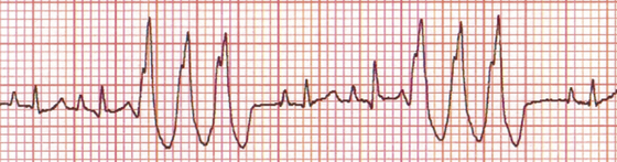
Figure 7-1 Lead II ECG tracing from a 7-year-old male, castrated Boxer with arrhythmogenic right ventricular cardiomyopathy. Ventricular premature beats with left bundle branch block morphology are a common finding in dogs with this condition. 25 mm/sec; 0.5 cm/mV.

Chest Radiography
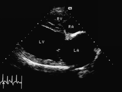
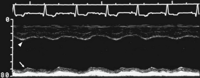
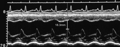
Echocardiography
• Echocardiography is widely used to quantify heart enlargement and systolic function. Routine echocardiography is not particularly sensitive in detecting early changes in occult disease, nor is it particularly helpful after a diagnosis of end-stage disease is made. As such, the utility of echocardiography increases as disease moves out of the occult phase to the overt clinical phase then declines again as the patient advances into end-stage disease. Early in the course of disease, many dogs possess normal echocardiographic examinations, despite having a significant numbers of ventricular arrhythmias.
Concomitant Abnormalities in Moderate or Severe Dilated Cardiomyopathy
• Azotemia is commonly detected in dogs that are receiving diuretic therapy and is typically prerenal in nature.
• More severe azotemia (blood urea nitrogen > 80 mg/dl and creatine > 3.0 mg/dl) can contribute to patient morbidity and may require reduction of angiotensin-converting enzyme (ACE) inhibitor and diuretic dose or parenteral fluid supplementation.


