9 Breeding Management
Most bitches are fairly consistent from one cycle to the next.
I. PREBREEDING EXAMINATION
If the goal of breeding is to produce puppies of higher quality than the parents, then prebreeding examination of the dam and sire is crucial to success. Both dogs should be evaluated for genetic disorders in their breed (see Chapter 29). Regardless of type of breeding intended (natural service versus artificial insemination) both the stud and dam should be tested for canine brucellosis (see Chapter 20). Genetic testing and brucellosis testing should be performed well in advance of the desired breeding to ensure that all tests can be completed before breeding occurs.
Both dogs also should undergo a complete physical examination to identify abnormalities impacting the health of the adult dogs, disease conditions that could affect the pups, and physical abnormalities that may preclude breeding or whelping. Physical examination and semen evaluation of the male should be performed at least 2 months before breeding if there is a concern about semen quality. After any insult to the testes, it takes at least 2 months to see improvement in semen quality. Males that are used regularly for breeding may benefit from consistent physical examination and semen evaluation every 6 months. Physical examination of bitches before breeding should include a digital vaginal examination to assess for the presence of a vaginal anomaly that may preclude natural service or whelping (see Chapter 17). If a stricture is palpated when the bitch is not in heat, the vagina should be reevaluated when the bitch is in heat. Under the hormonal influence of heat, vaginal tissues become more elastic and the stricture may relax sufficiently to allow natural breeding.
The owner of the stud dog may require vaginal culture of the bitch before mating. It must be recognized that the vagina is not sterile (Table 9-1). Semen is not sterile either, so if the owner of the male insists on a culture of the bitch, it is not untoward for the owner of the bitch to insist on a culture of the male. With either gender, a positive culture result does not mean anything in and of itself. A culture result can be considered significant only if there is moderate to heavy growth of one or two organisms. Mycoplasma culture is especially difficult to interpret because many laboratories will not give an idea of quantity when growth is reported. To treat all bitches with antibiotics when bred is not beneficial to the bitch or to the general population. It encourages development of bacteria that are resistant to antibiotics and, when therapy is concluded, allows a rebound of growth of organisms within the vagina.
Table 9-1 Normal Bacterial Flora of the Vagina of the Bitch
| Escherichia coli | Klebsiella spp. |
|---|---|
| Streptococcus spp. | Haemophilus spp. |
| Pasteurella spp. | Moraxella spp. |
| Staphylococcus spp. | Flavobacterium |
| Bacillus spp. | Pseudomonas spp. |
| Enterococcus spp. | Mycoplasma spp. |
| Proteus spp. |
If vaginal culture is to be performed before breeding, the following principles should be followed:
II. VAGINAL CYTOLOGY
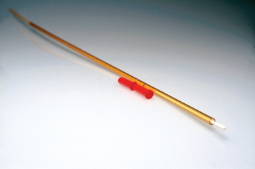
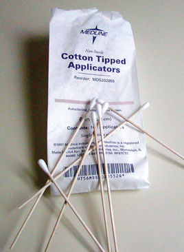
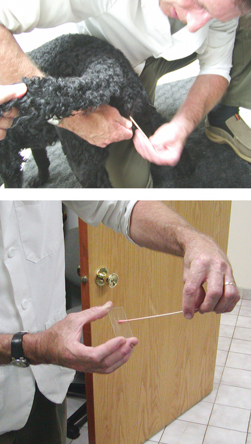
Cells are easily collected from the vagina of the bitch. Because the vagina is not sterile, a nonsterile swab can be used (Figure 9-2). The swab is moistened with water and introduced into the vaginal vault, cells are gently collected from the ceiling of the vagina, and the swab is withdrawn. The swab is rolled over a glass slide, and the slide is allowed to air-dry. The slide is stained and examined under the light microscope (Figure 9-3). Although individuals may interpret the types of cells present differently, there is no significant variation in management recommended for that dog. Still it is best to have all the vaginal cytology slides for a given bitch during a given estrus read by one person whenever possible.
Under the influence of the hormone estrogen, the cells of the vagina are stimulated to divide. As they divide and move farther and farther from the blood supply, the cells nearest the lumen, or opening, of the vagina die (Figure 9-4). These dead cells are called cornified or keratinized cells. As the bitch starts to secrete significant amounts of estrogen during proestrus, the population of collected cells changes. Evaluation of the percentage of cornified cells present on a vaginal swab gives a ballpark idea of what is happening in the dog’s cycle. Vaginal cytology alone cannot be used to determine optimal breeding day in the bitch.
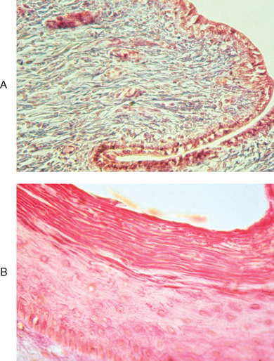
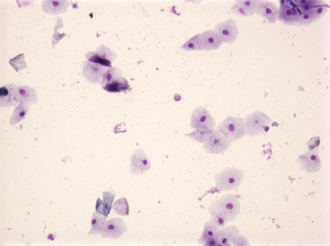
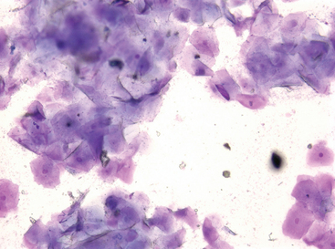
During early proestrus, most of the vaginal cells will be noncornified. These cells are round and healthy, with a healthy nucleus, and set in “nests” next to each other such that you easily can count the number of cells present. White blood cells may be present (Figure 9-5). As proestrus progresses, the percentage of cornified cells increases until in late proestrus virtually all the cells are cornified. White blood cells no longer appear in normal bitches at this stage of the cycle (Figure 9-6).
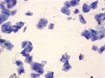
Estrous cytology is defined as completely cornified, with greater than half of the cells appearing to have no nucleus. The nucleus is there but is dead and does not take up stain. These cornified cells lie in clumps such that individual cells are difficult to identify. White blood cells are not present. Red blood cells may be identified. Bacteria commonly adhere to the surface of the cornified cells (Figure 9-7).
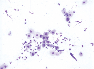
On the first day of diestrus, the cornified cells are abruptly shed from the vagina. The vaginal epithelial cell population abruptly returns to being noncornified. Many white blood cells are present in the first day or two of diestrus (Figure 9-8). This abrupt change from cornification to noncornification occurs quite consistently 6 days after ovulation.
< div class='tao-gold-member'>
Stay updated, free articles. Join our Telegram channel

Full access? Get Clinical Tree


