Blood vascular system

Chapter 7 Blood vascular system


KEY POINTS


The blood vascular system is made up of four parts:


Blood


Blood is a highly specialised fluid connective tissue (see Ch. 2), consisting of several types of cell suspended in a liquid medium called plasma. Blood makes up about 7% of the total body weight and has a pH of about 7.4. (7.35–7.45).


Functions


Blood has many functions within the body but they can be broadly divided into two groups: transport and regulation.


Regulation


Blood plays a vital role in homeostasis by regulating:


Composition of blood


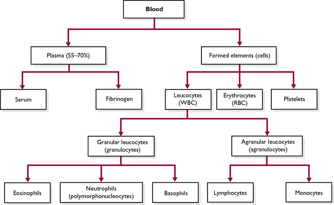
Blood is a red fluid that is carried by the blood vessels of the circulatory system. It is composed of a fluid part, the plasma, and a solid part, the blood cells (Fig. 7.1). Plasma forms part of extracellular fluid (ECF) (see Ch. 1). Each constituent of the blood plays a specific part in the overall function of blood.


Plasma


Plasma is the liquid part of the blood that separates out when a blood sample is spun in a centrifuge. The main constituent is water (about 90%) in which are a number of dissolved substances being transported from one part of the body to another. These include carbon dioxide in solution, nutrients such as amino acids, glucose and fatty acids, waste materials such as urea, hormones, enzymes, antibodies and antigens.


In addition to these, plasma contains:


Albumin, fibrinogen and prothrombin are produced by the liver, but the immunoglobulins are produced by the cells of the immune system.


Erythrocytes (or red blood cells)


Erythrocytes are the most numerous blood cell – there are about 6–8 million per cubic millilitre of blood (Fig. 7.2). Their function is to transport oxygen and a small proportion of carbon dioxide around the body (most carbon dioxide is carried in solution in the plasma).
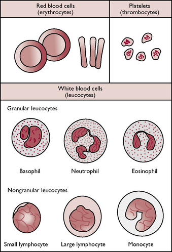


Mature erythrocytes are biconcave circular discs about 7 μm in diameter. Erythrocytes contain a red pigment called haemoglobin, which is a complex protein containing iron. They are the only cells in the body without a nucleus, which allows a greater amount of haemoglobin to be packed into a relatively small cell. Erythrocytes are surrounded by a thin, flexible cell membrane, which enables them to squeeze through capillaries. Their shape and thin cell membrane gives them a large surface area for gaseous exchange and allows oxygen to diffuse across into the cell, where it combines with the haemoglobin to form oxyhaemoglobin.


Erythrocytes are formed from undifferentiated stem cells within the bone marrow by a process known as erythropoiesis. The stem cells change into erythroblasts, which have a nucleus. The cell begins to acquire haemoglobin and its nucleus shrinks – it is now known as a normoblast. As the cell develops further it becomes a reticulocyte, at which point the nucleus consists only of fine threads in the cytoplasm known as Howell–Joly bodies. Eventually, the nucleus disappears and the mature erythrocyte is released into the circulation. This process takes 4–7 days.


If there is a shortage of erythrocytes, e.g. acute haemorrhage or iron-deficiency anaemia, reticulocytes are also released into the circulation to help make up the deficit. These can be seen on a blood smear stained with methylene blue, which is a specific stain for reticulocytes.


A circulating erythrocyte has a lifespan of about 120 days, after which it is destroyed in the spleen or lymph nodes. The iron from the haemoglobin is recycled back to the bone marrow and the remainder is converted by the liver into the bile pigment bilirubin and excreted in bile.


The production of red blood cells is controlled by a hormone called erythropoietin (see Ch. 6), which is released by cells in the kidney in response to low oxygen levels in the tissues. Erythropoietin stimulates the stem cells in the bone marrow to produce more erythrocytes.


Leucocytes (or white blood cells)


Leucocytes are much less numerous than red blood cells and the cells contain nuclei. Leucocytes can be classified as either granulocytes or agranulocytes depending upon whether or not they have visible granules in their cytoplasm when stained and viewed under a microscope (Fig. 7.2). The function of leucocytes is to defend the body against infection.


Granulocytes


This type of leucocyte is produced within the bone marrow and they make up approximately 70% of all leucocytes. They have granules within their cytoplasm and have a segmented or lobed nucleus which can vary in shape. They are referred to as polymorphonucleocytes or PMNs (meaning many-shaped nuclei). They can be further classified according to the type of stain they take up, i.e. neutral, basic or acidic. There are three types of granulocyte:


Neutrophils – take up neutral dyes and the granules stain purple (Fig. 7.2). Immature neutrophils have a nucleus that looks like a curved band and are known as band cells. Neutrophils are the most abundant of the leucocytes, forming about 90% of all granulocytes. They are able to move through the endothelial lining of the blood vessels into the surrounding tissues and engulf invading bacteria and cell debris by phagocytosis, thus helping to fight disease. A neutrophilia or raised numbers of neutrophils indicates the presence of an infective process while a neutropenia or lack of white cells may be characteristic of certain viral infections.

Eosinophils – these take up acidic dye and the granules in their cytoplasm stain red (Fig. 7.2). They are involved in the regulation of the allergic and inflammatory processes and secrete enzymes that inactivate histamine. Eosinophils play a major role in controlling parasitic infestation. An eosinophilia or raised numbers of eosinophils occurs in response to parasitic infestation.

Basophils – these take up basic or alkaline dyes and the granules in the cytoplasm stain blue (Fig. 7.2). Basophils secrete histamine, which increases inflammation, and heparin, which is a natural anticoagulant preventing the formation of unnecessary blood clots. Basophils are present in very small numbers in normal blood.


Agranulocytes


Agranulocytes have a clear cytoplasm. There are two types:


Lymphocytes – this is the second most common type of white blood cell, forming 80% of all agranulocytes (Fig. 7.2). Lymphocytes are the main cell type of the immune system and are formed in lymphoid tissue although they originate from stem cells in the bone marrow. Lymphocytes are responsible for the specific immune response, and there are two different types: the B lymphocytes, which produce antibodies and are involved in humoral immunity, and the T lymphocytes, which are involved in the cellular immune response.

Monocytes – these have a horseshoe-shaped nucleus and are the largest of the leucocytes, although they are only present in small numbers (Fig. 7.2). They are phagocytic cells and when they migrate to the tissues they mature and become known as macrophages.


Stay updated, free articles. Join our Telegram channel

Jul 18, 2016 | Posted by in PHARMACOLOGY, TOXICOLOGY & THERAPEUTICS | Comments Off on Blood vascular system

Full access? Get Clinical Tree

Get Clinical Tree app for offline access