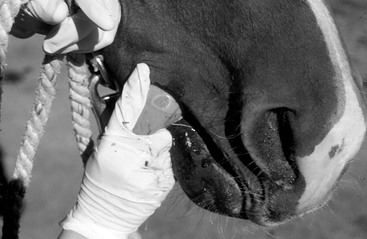
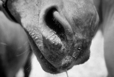
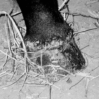
Brian J. McCluskey Vesicular stomatitis (VS) is a viral disease of horses, cattle, and other livestock and naturally occurs exclusively in the Western hemisphere. The disease is characterized by the appearance of short-lived vesicles that become ulcerations and erosions, especially on the tongue, oral and nasal mucosae, and coronary bands. The disease is caused by infection with either of two serotypes of vesicular stomatitis virus (VSV): VSV Indiana (VS-IN) or VSV New Jersey (VS-NJ). The prevalence rates of infection (as determined by seroconversion rates) and of clinical disease in horses vary greatly among individual premises during outbreaks. Disease may be apparent in animals at one site, but animals on neighboring premises remain clinically normal. Approximately 30% of infected animals on an affected property develop clinical signs. The prevalence of clinical infection in horses housed on pasture appears to be higher. This may be a result of factors affecting either pathogenicity of the virus (such as differences in mode of transmission) or susceptibility of the horses (such as differences in mucosal abrasions). After virus gains access to the animal host, virus multiplication is thought to remain localized in the epithelium, because viremia has not been detected in infected animals. After exposure, vesicles form in approximately 1 to 3 days. Lesions usually are healed in 7 to 14 days. Viral particles are present in high concentrations in vesicles and are shed from active lesions. Virus shedding ceases by 6 to 7 days after exposure. Vesicular stomatitis is clinically indistinguishable from foot and mouth disease (FMD) and is therefore a critical consideration in FMD control programs in the United States and worldwide. Likely as a result of this similarity (although FMD does not occur in equids), the United States is obligated to report confirmed findings of VS to the World Animal Health Organization (OIE). When VS is confirmed in the United States, strict transport embargoes are initiated by nonaffected states and most foreign countries, and movement of livestock is hindered by increased regulatory surveillance and quarantine of affected premises. The classic clinical sign of VS is the appearance of vesicular lesions on mucous membranes, with the oral cavity and nasal mucosa, tongue, and lips most commonly affected (Figures 127-1 to 127-3). Lesions may also appear on the mammary glands or external genitalia and coronary bands, and lesions on the ears and face have also been observed. As many as 70% of infected animals may show no overt signs of disease, and some infected animals may have only a few days of mild depression. After 1 to 3 days of incubation, blanched areas appear, which then develop into vesicles. Animals are generally febrile during this vesicular phase. There may be extensive mucosal necrosis and sloughing, especially of the dorsal surface of the tongue. Lesions generally resolve completely in 10 to 14 days. Serologic tests are available for detection of serum antibodies against VSV. A competitive enzyme-linked immunosorbent assay (cELISA) is used as a screening test. This test is rapid and detects both immunoglobulin M (IgM) and IgG. Serum neutralization (SN) and complement fixation (CF) tests are also used. Antibody can be detected within 5 to 8 days after infection. Both the cELISA and SN tests detect serum IgG, so antibody may be detectable in the serum for 1 to 3 years, making the interpretation of titers in a single serum sample difficult. A high CF titer in a single serum sample is more diagnostic of recent infection. A rising SN or CF titer is therefore required to definitively diagnose recent VSV infections. Virus isolation may be used to detect live virus in epithelial tags, swabs, or biopsy specimens from active lesions. Virus isolation is unrewarding after lesions have begun to resolve. Clinical vesicular stomatitis is typically short lived and self-limited. In most cases, no specific treatment is indicated, and horses recover uneventfully in 1 to 2 weeks. Even in severe cases, only supportive care and prevention of secondary complications are required until lesions resolve. Secondary bacterial infections can be minimized by frequent cleansing of lesions with common mild antiseptics or application of topical antimicrobials. Cachexia can be avoided or remedied by providing palatable feeds, such as grain or complete feed pellets softened with water. Providing fluids or feed through a stomach tube would be necessary only in horses that are severely anorexic. Rarely, dehydration becomes severe enough to necessitate intravenous fluid support. Corrective shoeing might be required if hoof wall deformities developing secondary to coronary band lesions become sufficiently severe to interfere with normal function. Vesicular stomatitis is a zoonotic disease, and good biosecurity practices, such as wearing disposable protective gloves and frequent handwashing, are recommended. Implementation of common management practices can reduce the risk for spread of this disease among and within premises. During regional outbreaks, all new arrivals to a facility or property should be considered suspect and should be quarantined for 3 to 5 days. No equipment, including feeders, waterers, salt blocks, brushes, and tack, should be shared among affected (or suspect) and nonaffected animals, and equipment and areas should be disinfected before noninfected animals are introduced. The viruses are easily inactivated with 1% formalin, 10% sodium hypochlorite, and other commonly used disinfectants. Vesicular stomatitis viruses are arthropod borne, so minimizing insect contact with livestock by stabling horses during periods of increased insect activity, using insect repellants on horses, controlling insects in areas where horses are housed, and moving animals away from pastures containing waterways during the summer months may help control disease spread. No vaccines are commercially available to prevent VSV infections in the United States.
Blistering Mucosal Diseases
Infectious Causes
Vesicular Stomatitis
Clinical Signs
Diagnosis
Treatment and Prevention
![]()
Stay updated, free articles. Join our Telegram channel

Full access? Get Clinical Tree


