Assessment of Acute-Onset, Severe Lameness

Chapter 13 Assessment of Acute-Onset, Severe Lameness


Field Diagnosis of the Injured Horse


The assessment of an acutely lame horse presents a challenge in diagnosis and in dealing with the people associated with the horse, particularly if lameness occurs at a competition. The horse may have fallen and been lame immediately or may have pulled up lame, and the veterinary surgeon may be called to examine the horse on course in full view of the public.


Ideally the horse should be transported to an examination area for comprehensive evaluation, but the veterinary surgeon must establish whether the injured limb requires support before the horse is moved. Although the horse may be very lame, establishing a definitive diagnosis for the cause of the lameness at this stage may be difficult.


This may surprise riders, trainers, and owners, and maintaining their confidence in what is an emotionally charged situation can be quite difficult. If a fracture is suspected, pressure to destroy the horse humanely without delay may be felt. Although some fractures are catastrophic and merit immediate destruction of the horse on humane grounds (e.g., a spiral fracture of the humerus), other serious fractures can be repaired. Therefore as much information as possible about the site of the fracture and its configuration should be obtained before a decision is made. The limb should be supported appropriately before the horse is moved for radiographic examination. If a horse must be destroyed on humane grounds at a competition, this should be done off the course.


Although a diagnosis may be obvious in some horses immediately after the onset of lameness, the veterinarian must recognize that severe lameness may occur without an evident cause. Serial reexaminations over the following hours or days may be required before a diagnosis can be reached. Sometimes the lameness resolves spontaneously within 12 to 18 hours and its cause is never established.


The clinician must be aware of the most common causes of acute-onset, severe lameness, which include the following:


When lameness occurs during training or competition, a spectrum of other injuries must be considered. However, a history of acute-onset, severe lameness during exercise must not mislead the clinician into thinking that lameness must be caused by internal or external trauma associated with exercise. Lameness may still be caused by pain from a subsolar abscess.


This chapter describes a systematic approach to management of a horse with sudden-onset, severe lameness and focuses particularly on injuries that occur during work.


Assessment


Medical History


While performing an initial visual appraisal of the horse, establishing a history is useful. The examiner must determine the following:


The clinician should also be aware of common injuries in the discipline in which the horse is competing.


The horse may be distressed because of the severity of pain and excited because of the atmosphere of a competition and thus difficult to restrain and examine adequately. Sedation with romifidine or detomidine, with or without butorphanol, may be necessary to facilitate examination of the horse. A horse with an acute hindlimb muscle tear or hemorrhage may show evidence of pain mimicking signs of colic.


The horse’s posture should be observed while it stands still and walks a few steps. If the horse bears weight only on the toe, it may be inapparent that the horse has lost some support of the fetlock because of rupture of the superficial digital flexor tendon (SDFT) in the metacarpal region or at the musculotendonous junction in the antebrachium, unless it walks a few steps.


Limb Examination


The veterinarian should establish whether the horse is able and willing to bear weight on the limb, bearing in mind that after a fall a neurological component may contribute to the lameness, in addition to the pain. The horse’s demeanor should be assessed; the degree of pain and distress usually but not invariably reflects the severity of the injury. The horse may be greatly distressed, shifting weight constantly between limbs, and may be reluctant to move. Reluctance to move may be caused by a bilateral problem (e.g., bilateral severe superficial digital flexor [SDF] tendonitis) or a more generalized problem such as equine rhabdomyolysis (tying up).


The horse should be carefully appraised visually to identify areas of swelling or a laceration. If the horse’s limbs are covered in mud or grease (commonly applied to the limbs during the speed and endurance phase of a Three Day Event), this should be washed off before one proceeds with the evaluation. Boots, bandages, and the saddle and martingale should also be removed. Temporary studs in the shoes should be removed because they may be more difficult to remove later if the injury is severe.


The horse may be obviously lame on a hindlimb or forelimb, but this may mask a similar, less severe injury in a contralateral limb or a different injury; therefore all limbs should be assessed carefully. For example, a racehorse may develop a lateral condylar fracture of the third metacarpal bone in one limb and SDF tendonitis in the contralateral limb. Although the former injury results in a more severe lameness, the latter may be more important to the horse’s long-term prognosis.


Occasionally, forelimb and hindlimb lameness are concurrent. Each limb should be palpated systemically with the horse bearing weight and not bearing weight. The examiner should pay careful attention to heat, swelling, abnormal muscle texture, pain on firm pressure, pain induced by manipulation of a joint, restriction of flexibility of a joint, an abnormal range of motion of the joint, audible or palpable crepitus, and the intensity of the digital pulse amplitudes.


The position of the shoe should be assessed carefully. A shoe that has moved slightly may result in nail bind. Hoof testers should be systemically applied across the wall and sole, gently at first and then firmly. Percussion should also be applied to the sole of the foot with the limb picked up and to the wall with the limb bearing weight. The clinician should not forget that if the sole is very hard, eliciting pain with hoof testers may not be possible, despite the presence of a subsolar abscess.


The limbs should be carefully assessed for lacerations. Serious damage to underlying structures may have occurred if the laceration was sustained while the horse was moving at speed, and the position of the laceration and the site of damage to underlying structures may not coincide.


Shoulder and Chest


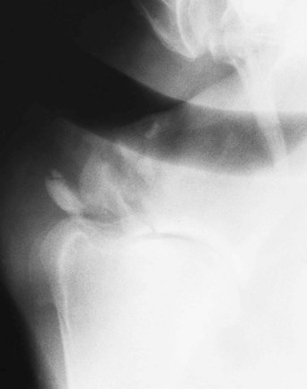
Injuries to the shoulder region usually result from a fall or collision, which may result in severe bruising only or a fracture. A fracture of the supraglenoid tubercle of the scapula results in severe lameness (Figure 13-1). Slight soft tissue swelling may develop, usually without audible or palpable crepitus, and pain on palpation may be difficult to differentiate from that caused by severe bruising alone. Articular fractures of the scapula may be associated with audible crepitus on manipulation of the limb. Fractures of the body of the scapula or the humerus are usually associated with severe lameness, soft tissue swelling, and pain in that area.


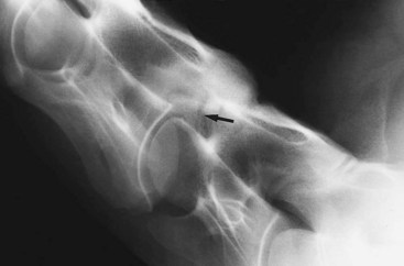
After collision with a fixed object, or occasionally a fall, the scapulohumeral joint may become luxated or subluxated, with or without a fracture of the glenoid cavity of the scapula. The horse bears weight on the limb reluctantly, soft tissue swelling develops rapidly, and the distal aspect of the scapular spine may become more difficult to palpate. The limb may appear straighter than usual. A collision also may result in collateral instability of the shoulder, so-called shoulder slip, usually caused by trauma to nerves of the brachial plexus. The horse may have pain-related lameness because of bruising, together with mechanical lameness caused by neurological dysfunction (Figure 13-2).


Although major fractures of the scapula and humerus are usually readily evident by clinical signs, most other shoulder injuries require radiographic and sometimes ultrasonographic examination for a diagnosis to be reached.


Pectoral muscle tears may result in similar clinical signs, with severe lameness and distress. Repeated clinical examinations may reveal the site of muscle rupture, with increasing evidence of hemorrhage, inflammatory effusion, and edema.

Stay updated, free articles. Join our Telegram channel

Jun 4, 2016 | Posted by in EQUINE MEDICINE | Comments Off on Assessment of Acute-Onset, Severe Lameness

Full access? Get Clinical Tree

Get Clinical Tree app for offline access