Arthrology


Arthrology


General


Articulations, or joints (articulationes [juncturae] ossium), are formed when two or more bones are united by fibrous, elastic, or cartilaginous tissue or by a combination of these tissues. Three main groups are recognized and named according to their most characteristic structural features. Where little movement is required, the union is short, direct, and often transitory. A fibrous joint (junctura fibrosa), formerly known as a synarthrosis, is one of this nature. Such joints include syndesmoses, sutures, and gomphoses. A cartilaginous joint (junctura cartilaginea), formerly known as an amphiarthrosis, permits only limited movement, such as compression or stretching. A synovial joint (junctura synovialis) formerly known as a diarthrosis or true joint, facilitates mobility. The studies of Kadletz (1932) provide detailed information on the arthrology of the dog, and the well-documented work of Barnett et al. (1961) discusses the structure and mechanics of synovial joints in considerable detail.


The term syndesmologia was used in the Basel Nomina Anatomica (BNA) of 1895 for the joints and ligaments. This was changed to arthrology in the Birmingham Revision of 1933 and back to the original in Paris in 1955.


At the Tokyo meeting of the International Nomenclature Committee, arthrologia was adapted as the most appropriate heading and articulatio replaced junctura. The sixth edition of Nomina Anatomica (1989), retained arthrologia and articulatio. It should be noted that the discarded original term, syndesmologia, for all joints is similar sounding to the term syndesmosis, which is used to denote one type of fibrous joint.


Nomina Anatomica Veterinaria (1983) adopted articulatio for all joints—fibrous, cartilaginous, and synovial. The term articulationes synoviales replaces the former terms diarthrosis and articulus. This terminology was retained in the fifth edition of the Nomina Anatomica Veterinaria in 2005.


Fibrous Joints


A syndesmosis is a fibrous joint with a considerable amount of intervening connective tissue. The attachment of the hyoid apparatus to the petrous part of the temporal bone is an example of a syndesmosis.


A suture (sutura) is a fibrous joint of the type that is confined largely to the flat bones of the skull. Depending on the shape of the apposed edges, sutures are further divided into (1) serrated suture (sutura serrata), one that articulates by means of reciprocally alternating processes and depressions; (2) squamous suture (sutura squamosa), one that articulates by overlapping of reciprocally beveled edges; (3) plane suture (sutura plana), one in which the bones meet at an essentially right-angled edge or surface; and (4) foliate suture (sutura foliata), one in which the edge of one bone fits into a fissure or recess of an adjacent bone. Serrate sutures are found where stable noncompressible joints are needed, such as the parietooccipital and the interparietal unions. Where a slight degree of compressibility is advantageous, such as is required in the fetal cranium at birth, squamous sutures are found. Similarly, the frontonasal and frontomaxillary squamous sutures allow enough movement to absorb the shock of a blow that might otherwise fracture the bones of the face. Examples of plane sutures are those of the ethmoid and those between most of the bones of the face. Where extreme stability is desirable, foliate sutures are formed. The best example of this type is the zygomaticomaxillary suture. The various fibrous sutures of the skull also permit growth to take place at the periphery of the bones. When uneven jagged edges of bones interlock in a fibrous joint, as occurs in several skull bones, it is called a schindylesis.


The implantation of a tooth in its alveolus by means of a fibrous union known as a gomphosis, or articulatio dentoalveolaris. This specialized type of fibrous joint is formed by the periodontal ligament (periodontium), which attaches the cementum of the tooth to the alveolar bone of the alveolus and permits slight movement.


Cartilaginous Joints


Many bones are united by cartilaginous joints, which are sometimes referred to as synchondroses. Unions of this type may be formed by hyaline cartilage, by fibrocartilage, or by a combination of the two, and they are subject to change with increasing age.


Hyaline cartilage joints, or primary joints, are usually temporary and represent persistent parts of the fetal skeleton or secondary cartilage of growing bones. The epiphysis of an immature long bone is united with the diaphysis by a cartilaginous physeal plate. When adult stature is reached, osseous fusion occurs and a joint no longer exists, although a slight physeal line may mark the union. This osseous union in some anatomic works is called a synostosis. Similar transitory hyaline cartilage joints are typical of the sphenooccipital synchondrosis or the union of an apophysis with the extremity (epiphysis) or body (diaphysis) of a long bone such as with the femoral trochanters or the ulnar olecranon tubercle. The humeral tubercles develop from the proximal epiphysis. Some hyaline cartilage joints, such as the costochondral junctions, remain throughout life.


Fibrocartilaginous joints, or secondary joints, are sometimes referred to as amphiarthroses. The best examples of such joints are those of the pelvic symphysis, the intermandibular articulation, sternebrae, and vertebral bodies. The fibrocartilage uniting these bones may have an intervening plate of hyaline cartilage at each end. Occasionally these joints may ossify, as do hyaline cartilage joints.


Synovial Joints


The synovial joints of the extremities permit the greatest degree of movement and are most commonly involved in dislocations. All synovial joints (articulationes synoviales) are characterized by a joint cavity (cavum articulare), a joint capsule (capsula articularis) including an outer fibrous layer and an inner synovial membrane, synovial fluid (synovia), and articular cartilage (cartilago articularis). Collateral ligaments are developed in the fibrous layer of the joint capsule. A few of the synovial joints have modifications of their joint capsules peculiar to the functions they perform and may possess intraarticular ligaments, menisci, fat pads, or synovial membrane projections in the form of plicae or villi. These are primarily developments of the fibrous membrane of the joint capsule.


The blood supply of synovial joints is provided by an arterial and venous network from parent trunks in the vicinity of the joint. The vessels supply the capsule and also the epiphyses bordering the joint. Around the articular margins, the blood vessels of the synovial membrane form anastomosing loops, referred to collectively as the circulus articularis vasculosus.


Lymphatic vessels are also present in synovial membranes and account for the rapid removal of some substances from the joint cavity.


The nerve supply of synovial joints is derived from cutaneous or muscular branches in the vicinity of the joint. Included in these articular nerves are proprioceptive fibers, nociceptor fibers and sympathetic visceral efferent and visceral afferent fibers related to vasomotor or vasosensory functions respectively. Some areas of the joint capsule are more richly innervated than others. Four types of joint receptors are present in most animal joints (Polacek, 1966; Zimny, 1988): (1) Ruffini-like receptors in the capsule, (2) Pacinian-like receptors in the capsule, (3) Golgi tendon organs in ligaments, and (4) free nerve endings. If a joint has intraarticular structures, they are usually innervated. The purpose of the innervation is proprioception and the recognition of angular movement; thus posture is very dependent on these endings. It is likely that the stifle joint with its many ligaments and menisci has the richest innervation of all joints. O’Connor and Mc-Connaughey (1978) and O’Connor (1976, 1984) found both Ruffini endings and Pacinian corpuscles in the menisci of the dog and cat. Sfameni (1902) found single or grouped nerve endings that arose from single axons in the dog and suggested the name Ruffini endings because they resembled those described in the skin by Ruffini in 1894. Gardner (1950) reviewed the morphologic and physiologic characteristics of joints in the human, including their innervation, and cites more than 500 references. Ansulayotin (1960) studied the nerves that supply the appendicular joints in the dog.


Structure of Synovial Joints


The joint capsule is composed of an inner synovial membrane and an outer fibrous membrane. The synovial membrane (membrana synovialis) is a vascular connective tissue that lines the inner surface of the capsule and is responsible for the production of synovial fluid. The synovial membrane does not cover the articular cartilage but blends with the periosteum as it reflects onto the bone. Joint capsules may arise postnatally if the need exists, and thus false joints often form following unreduced fractures. Synovial membrane covers all structures within a synovial joint except the articular cartilage and the contact surfaces of fibrocartilaginous plates. Synovial membrane also forms sleeves around intraarticular ligaments and covers muscles, tendons, nerves, and vessels if these cross the joint closely. Adipose tissue often fills the irregularities between articulating bones, and in some instances it is aspirated into or squeezed out of the joint as the surfaces of the articulating bones part or come together during movement. Fat in such locations is covered by synovial membrane. A synovial fold (plica synovialis) is an extension of the synovial membrane; such folds usually contain fat. Around the periphery of some synovial joints the synovial membrane is in the form of numerous processes, or synovial villi (villi synoviales). These are soft and velvety. The synovial membrane may extend beyond the fibrous layer and act as a bursa deep to a tendon or ligament, or may even form a synovial sheath.


The fibrous membrane (membrana fibrosa) of a joint capsule is composed mainly of white fibrous tissue containing yellow elastic fibers. It is also known as the capsular ligament. In most joints the ligaments are thickenings of the fibrous portion of the joint capsule. In some synovial joints the ligaments appear to be quite separate from the fibrous capsular ligament, such as the patellar ligament of the stifle joint. Maybe this is reason to consider the patellar ligament to be the tendon of insertion of the quadriceps muscle with a sesamoid (the patella) associated with this insertion. On the other hand, the patellar ligament can be considered to be a development of the fibrous layer of the stifle joint capsule along with the extensive fat pad associated with it. In those joints where great movement occurs in a single plane the fibrous membrane is usually thin and loose on the flexor and extensor surfaces, and thick on the sides of the bone that move the least. Such thickenings of the fibrous layer are known as collateral ligaments (ligg. collateralia) and are present to a greater or lesser degree in all hinge joints. The fibrous membrane attaches at the margin of the articular cartilage, or at most 3 cm from it, where it blends with the periosteum.


The synovial fluid (synovia) serves chiefly to lubricate the contact surfaces of synovial joints. In all cases these surfaces are hyaline cartilage or fibrocartilage. Fibrocartilage contains few blood vessels and nerves, and hyaline cartilage has neither. Therefore the synovial fluid serves the additional function of transporting nutrient material to the hyaline cartilage and removing the waste metabolites from it. Synovia also enables the wandering leukocytes to circulate in the joint cavity and phagocytize the products of the wear and tear of the articular cartilage. In many joints there is little, if any, free synovia. The average volume in the stifle joint of adult dogs of various sizes varies from 0.2 mL to 2 mL. The general health and condition of the dog has a marked influence on the amount of synovia present in the joints. Synovia is thought to be a dialysate, although mucin is probably produced by the fibroblasts of the synovial membrane (Davies, 1944). The chemical composition of synovia closely resembles that of tissue fluid. In addition to mucin, it contains salts, albumin, fat droplets, and cellular debris. The quantitative composition of synovia depends largely on the type of tissue underlying the surface fibroblasts and the degree of vascularity of this tissue. Because of its mucin content, the synovia forms a viscous capillary film on the articular cartilage.


The articular cartilage (cartilago articularis) is usually hyaline cartilage. It covers the articular surfaces of bones where its deepest part may be calcified. It contains no nerves or blood vessels, although it is capable of some regeneration after injury or partial removal (Bennett et al., 1932). It receives its nutrition from the synovia. The articular cartilage varies in thickness in different joints and in different parts of the same joint. It is thickest in young, healthy joints and in joints that bear considerable weight. Its thickness in any particular joint is in direct proportion to the weight borne by the joint, and it may atrophy from disuse. Healthy articular cartilage is translucent, with a bluish sheen. Elasticity and compressibility are necessary physical properties that it possesses. This resiliency guards against fracture of bone by absorbing shock.


A meniscus (meniscus articularis), or disc (discus articularis), is a complete or partial fibrocartilaginous plate that divides a joint cavity into two parts. The temporomandibular joint contains a thin, but complete, articular disc, and, because the capsular ligament attaches to the entire periphery of the disc, the joint cavity is completely divided into two parts. Two menisci are found in the stifle joint, and neither is complete, thus allowing all parts of the joint cavity to intercommunicate. Menisci have a small blood and nerve supply and are capable of regeneration (Dieterich, 1931). Their principal function, according to MacConaill (1932), is “to bring about the formation of wedge-shaped films of synovia in relation to the weight-transmitting parts of joints in movement.” An obvious function is the prevention of injury from concussion. The stifle and temporomandibular joints are the only synovial joints in the dog that possess menisci, or discs.


A ligament (ligamentum) is a band or a cord of nearly pure collagenous tissue that unites two or more bones. The term has also been used to designate remnants of fetal structures and relatively avascular narrow serous membrane connections. Ligaments, as used in this chapter, unite bone with bone. Tendons unite muscle with bone. Most ligaments are extraarticular but a few are intraarticular such as in the stifle and hip joints. They always develop initially within the fibrous layer of the joint capsule. The loss of intraarticular components of the joint capsule may result in what appear to be ligaments within a joint unassociated with a joint capsule. They are covered by synovial membrane. They are heaviest on the side of joints where the margins of the bones do not separate but glide on each other. Hinge joints with the greatest radii of movement have the longest ligaments. The ligaments often widen at their attached ends, where they blend with the periosteum. Histologically, ligaments are composed largely of long parallel or spiral collagenous fibers, but all possess some yellow elastic fibers also. The integrity of most joints is ensured by the ligaments, but in some (shoulder and hip) the heavy muscles that traverse the joints play a more important part in the function of that joint than do the ligaments. Such muscles and their tendons are sometimes spoken of as active ligaments. In hinge joints ligaments limit lateral mobility, and some (cruciate ligaments of the stifle joint) limit folding, opening, and sliding of the joint as well. In certain ball-and-socket synovial joints the sockets are deepened by ridges of dense fibrocartilage, known as glenoid lips (labia glenoidalia).


Classification of Synovial Joints


Synovial joints may be classified according to (1) the number of articulating surfaces involved, (2) the shape or form of the articular surfaces, or (3) the function of the joint (Barnett et al., 1961).


According to the number of articulating surfaces a joint is either simple (articulatio simplex) or compound (articulatio composita). A simple joint is formed by two articular surfaces within an articular capsule. When more than two articular surfaces are enclosed within the same capsule, the joint is compound.


The classification of synovial joints (Nomina Anatomica Veterinaria, 2005) is based on the shape or form of the articular surfaces. There are seven basic types:


A plane joint (articulatio plana) is one in which the articular surfaces are essentially flat. It permits a slight gliding movement. An example is the costotransverse joint.


A ball-and-socket joint (articulatio spheroidea) is formed by a convex hemispherical head that fits into a shallow glenoid cavity (shoulder joint) or into a deep cotyloid cavity (hip joint).


An ellipsoidal joint (articulatio ellipsoidea) is similar to a spheroidal joint. It is characterized by an elongation of one surface at a right angle to the other, forming an ellipse. The reciprocal convex (male) and concave (female) elongated surfaces of the antebrachiocarpal articulation form an ellipsoidal joint.


A hinge joint (ginglymus) permits flexion and extension with a limited degree of rotation. The most movable surface of a hinge joint is usually concave. An example is the elbow joint.


A condylar joint (articulatio condylaris) resembles a hinge joint in its movement but differs in structure. The surfaces of such a joint include rounded prominences, or condyles, that fit into reciprocal depressions or condyles on the adjacent bone, resulting in two articular surfaces usually included in one articular capsule. Examples of condylar joints include the temporomandibular joint and the stifle joint. The stifle joint is best classified as a complex condylar joint, because it possesses an intraarticular fibrocartilage that partially subdivides the intraarticular cavity.


A trochoid (articulatio trochoidea), or pivot joint, is one in which the chief movement is around a longitudinal axis through the bones forming the joint. The median atlantoaxial joint and the proximal radioulnar joint are examples of trochoid joints.


A saddle joint (articulatio sellaris) is characterized by opposed surfaces, each of which is convex in one direction and concave in the other, usually at right angles. When opposing joint surfaces are concavo-convex, the main movements are also in planes that meet at right angles. The tarsocrural or interphalangeal joints are examples of this type of articulation.


Movements of Synovial Joints


Joint movements that are brought about by the contraction of muscles that cross the joints are known as active movements. Those joint movements caused by gravity or secondarily by the movement of some other joint or by an external force are known as passive movements. Synovial joints are capable of diverse movements. Flexion, or folding, denotes moving two or more bones so that the angle between them becomes less than 180 degrees. Extension, or straightening, denotes movement by which the angle is increased to 180 degrees. It is readily seen that some joints, such as the metacarpophalangeal and metatarsophalangeal joints, are in a resting state of overextension. This is also called dorsal flexion. When an animal “humps up,” it flexes its vertebral column. Some parts of the vertebral column (the joints between the first few caudal vertebrae) are normally in a state of flexion, whereas others (the joints between the last few cervical vertebrae) are in a state of overextension. Flexion and extension occur in the sagittal plane unless the movement is specifically stated to be otherwise (right or left lateral flexion of the vertebral column). Adduction is the term applied to moving an extremity toward the median plane or a digit toward the axis of the limb. Abduction, or taking away, is the opposite movement. Circumduction occurs when an extremity follows in the curved plane of the surface of a cone. Rotation is the movement of a part around its long axis.


Ligaments and Joints of the Skull


Temporomandibular Joint


The temporomandibular joint (articulatio temporomandibularis) (Figs. 5-1 and 5-2) is a condylar joint that allows considerable sliding movement. The transversely elongated condyle of the mandible does not correspond entirely to the articular surface of the mandibular fossa of the temporal bone. A thin articular disc (discus articularis) lies between the cartilage-covered articular surface of the condyloid process of the mandible and the similarly covered mandibular fossa of the temporal bone.


The loose joint capsule extends from the articular cartilage of one bone to that of the other. On the temporal bone the capsular ligament also attaches to the retroarticular process. It attaches to the entire edge of the disc as it passes between the two bones. The joint cavity is thus completely divided into a dorsal compartment, between the disc and temporal bone, and a ventral compartment, between the disc and mandible. Laterally the fibrous part of the joint capsule is strengthened by fibrous strands to form the lateral ligament (lig. laterale). The lateral ligament becomes progressively tighter as the jaws open, and if one or the other is unduly lax owing to stretching or joint dysplasia, it is possible to dislocate the temporomandibular joint. Robins and Grandage (1977) described open-mouth jaw locking and its surgical correction in two Basset Hounds with temporomandibular joint dysplasia. Differential movement at the joints, when the jaws were opened widely, allowed locking of the coronoid process lateral to the zygomatic arch.


Vollmerhaus and Roos (1996) described transverse movement of the temporomandibular joint in 20 dogs of various breeds. This movement is important for mastication. Umphlet et al (1988) described the effect of hemimandibuloectomy on the joint.


Intermandibular Joint


The intermandibular articulation (articulatio intermandibularis) includes a small part formed by cartilage. This is the median synchondrosis (synchondrosis intermandibularis) uniting right and left mandibular bodies. The larger part of the articulation consists of connective tissue forming a suture (sutura intermandibularis). The opposed articular surfaces are interdigitated, and the fibrocartilage of the articulation may persist throughout life.


Scapino (1965, 1981) investigated the morphologic characteristics and function of the intermandibular articulation in the dog and other carnivores. He described four types of articulations, ranging from flexible to synostosed. He considers the dog to have a flexible joint that permits a moderate amount of independent movement of the mandibles and found this to be the most common type of union in carnivores. When the mandibles of such a joint are separated, the articular surfaces are flat or have low rugosities. A smooth area can be seen rostrodorsally, and the articular space is usually wider caudally than rostrally. The joint is characterized by a single fibrocartilage pad, cruciate ligaments, and a venous plexus. In the wolf and dog the articulation is not stiff, as in the lion and tiger, and is not synostosed, as in the badger and panda.


Joints of Auditory Ossicles


The joints of the auditory ossicles (articulationes ossiculorum auditus) allow for movement of the malleus, incus, and stapes (see Chapter 20). The head of the malleus articulates with the body of the incus via a synovial incudomallear joint (articulatio incudomallearis). The lenticular process of the long crus of the incus, with the head of the stapes, likewise forms a synovial joint, which is called the incudostapedial joint (articulatio incudostapedia). The footplate, or base, of the stapes attaches to the margin of the vestibular window (fenestra vestibuli) by means of a fibrous union (syndesmosis tympanostapedia).


The ligaments of the auditory ossicles (ligg. ossiculorum auditus) function to hold the ossicles in place and to limit their movement. Associated with the malleus (lig. mallei) is a short lateral ligament between the lateral process of the malleus and the tympanic notch, a dorsal ligament joining the head of the malleus to the roof of the epitympanic recess, and a short rostral ligament connecting the rostral process of the malleus to the osseous tympanic ring. The body of the incus is attached to the roof of the epitympanic recess by a dorsal ligament, and the short crus of the incus is attached to the fossa incudis by a caudal ligament. The base of the stapes is attached to the margin of the vestibular window by an annular ligament (lig. annulare stapedis).


Sutures of the Skull


The sutures of the skull (suturae capitis) are described in the discussion of the individual bones of the skull in Chapter 4. The name of each bone in the following list is followed by the names of the sutures in which it participates.


Occipital Bone


Parietal Bone


Frontal Bone


Sphenoid Bone


Temporal Bone


Ethmoid Bone


Incisive Bone


Nasal Bone


Maxilla


Zygomatic Bone


Palatine Bone


Lacrimal Bone


Pterygoid Bone


Vomer


Ligaments and Joints of the Vertebral Column


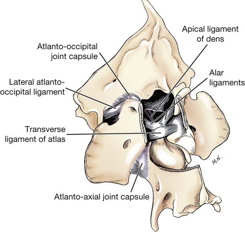
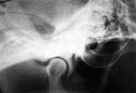
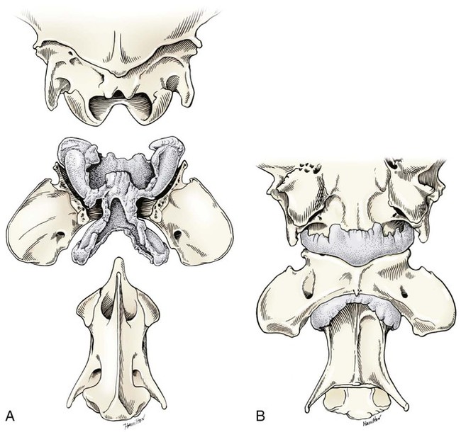
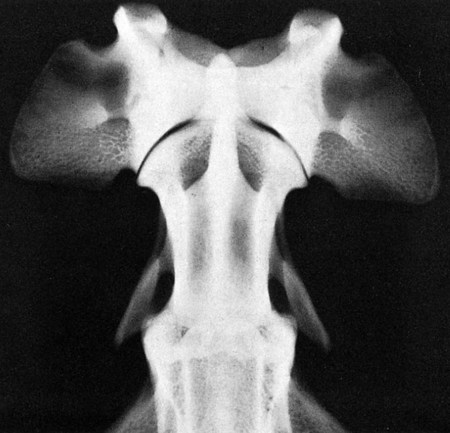
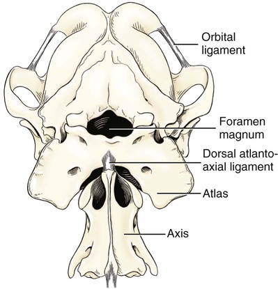
Atlantooccipital Articulation


There is a common joint cavity formed by the articulations of the occipital condyles with the atlas, and the atlas with the axis. By means of silicone casts this cavity (Fig. 5-3) has been studied and described as the “composite occipito-atlas-axis joint cavity” by Watson et al. (1986). A cast of the cavity resembles the shape of an “hourglass” with the ends removed or “popeye” holding up the head. It appears to be a composite of five synovial joints: right and left atlantooccipital joints, a median joint cavity between the ventral articular surface of the dens and the dorsal surface of the ventral arch of the atlas, and right and left atlantoaxial joints. The synovial bursa between the transverse atlantal ligament and the dens does not communicate with the common joint cavity.


The atlantooccipital joint (articulatio atlantooccipitalis) is formed by the dorsolaterally extending occipital condyles and the corresponding concave cranial articular fovea of the atlas. The spacious joint capsule (capsula articularis) on each side attaches to the margins of the opposed articular surfaces. Ventromedially the two sides are joined so that an undivided U-shaped joint cavity is formed. The atlantooccipital joint cavity communicates with the atlantoaxial joint cavity along the dens. The dorsal and ventral atlantooccipital membranes reinforce the joint capsule at their respective locations.


The dorsal atlantooccipital membrane (membrana atlantooccipitalis dorsalis) extends between the dorsal edge of the foramen magnum and the cranial border of the dorsal arch of the atlas. Two oblique straplike thickenings, approximately 8 mm wide, arise on each side of the notch of the squama occipitalis, diverge as they run caudally, and attach to the dorsolateral parts of the atlas. In the triangular space formed by these bands, punctures are made for the removal of cerebrospinal fluid from the cerebellomedullary cistern.


The ventral atlantooccipital membrane (membrana atlantooccipitalis ventralis) and its synovial layer form the uniformly thin joint capsule located between the ventral edge of the foramen magnum and the ventral arch of the atlas.


The lateral ligament (lig. laterale) of the atlantooccipital joint (see Fig. 5-5) runs from the lateral part of the dorsal arch of the atlas to the paracondylar process of the occipital bone. Its course is cranioventrolateral, and its caudal attachment is narrower than its cranial one. Another small ligament runs from each side of the inner surface of the lateral part of the ventral arch of the atlas to the lateral part of the foramen magnum. Ventral and medial to these ligaments the unpaired joint cavities between the skull and the atlas and between the atlas and the axis freely communicate.


Atlantoaxial Articulation


The atlantoaxial joint (articulatio atlantoaxialis) (see Figs. 5-3 to 5-6) is a pivot joint that permits the head and atlas to rotate around a longitudinal axis. The joint capsule is loose and uniformly thin as it extends from the dorsal part of the cranial articular surface of one side of the axis to a like place on the opposite side. Cranially it attaches to the caudal margins of the caudal articular foveae and ventral arch of the atlas. The fibrous layer of the joint capsule extends from right to left between the dorsal arch of the atlas and the arch of the axis. This is the dorsal atlantoaxial membrane, or membrana tectoria.


The apical ligament of the dens (lig. apicis dentis) (see Fig. 5-5) leaves the apex of the dens and passes straight cranially to the basioccipital bone at the ventral part of the foramen magnum. The apical ligament represents a remnant of the notochord. The two alar ligaments (ligg. alaria) are wider and heavier than the apical ligament. They attach to the dens on either side of the apical ligament and diverge from each other to attach to the occipital bone medial to the caudal parts of the occipital condyles. The transverse atlantal ligament (lig. transversum atlantis) is a thick ligament that connects one side of the ventral arch of the atlas to the other. It crosses dorsal to the dens and functions to hold this process against the ventral arch of the atlas. A spacious bursa exists between the ventral surface of the ligament and the dens.


Atlantoaxial subluxation with absence of the dens has been reported frequently, particularly in toy breeds, and has been ascribed to either congenital developmental or degenerative causes. Injury may result in a fracture of the dens. In almost all instances there is a tilting or dorsal displacement of the axis into the vertebral canal, with resultant compression of the spinal cord (Cook & Oliver, 1981; Oliver & Lewis, 1973).


Other Synovial Joints of the Vertebral Column


The synovial joints of the vertebral column caudal to the axis are those that appear in pairs between the articular processes of contiguous vertebrae (articulations processuum articularum), also known as juncturae zygapophyseales, and the joints between the ribs and the vertebrae (articulationes costovertebrales). The articular process joint capsules (capsular articularis) are most voluminous in the cervical region and at the base of the tail, where the greatest degrees of movement occur. The articular processes of all vertebrae cranial to the tenth thoracic are in nearly a dorsal plane so that the cranial articular processes face dorsally and the caudal articular processes face ventrally. At the tenth thoracic vertebra the direction of the articular processes changes. From the articulation between the tenth and eleventh thoracic vertebrae and through all the lumbar vertebral articulations there is essentially a sagittal interlocking of the cranial and caudal articular processes. The caudal articular processes of this segment face laterally, and the cranial articular processes face medially.


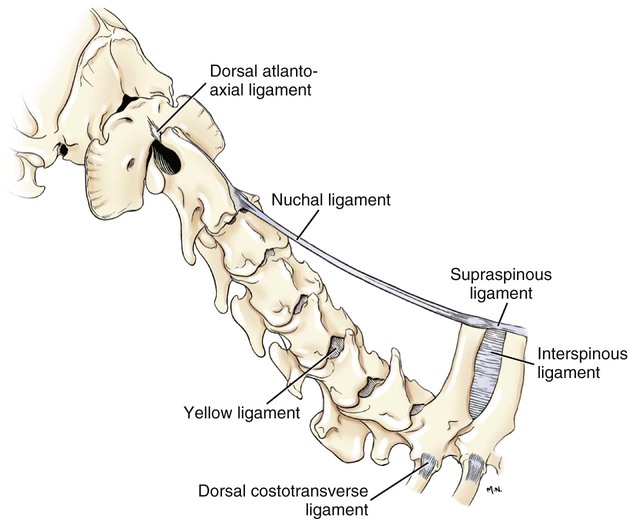
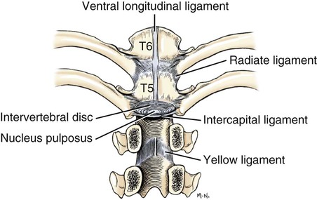
Long Ligaments of the Vertebral Column


The nuchal ligament (lig. nuchae) (Fig. 5-7) is composed of longitudinal yellow elastic fibers that attach cranially to the caudal part of the large spinous process of the axis. It extends caudally to the dorsal extremity of the spinous process of the first thoracic vertebra. It is a laterally compressed, paired band that lies between the medial surfaces of the mm. semispinales capiti. The yellow nature of the nuchal ligament continues caudally in the supraspinous ligament to the tenth thoracic spinous process (Baum & Zietzschmann, 1936).


The supraspinous ligament (lig. supraspinale) (see Figs. 5-12 and 5-7) extends from the spinous process of the first thoracic vertebra caudally to the third caudal vertebra. It is a thick band especially in the thoracic region, where it attaches to the apices of the spines as it passes from one to another. Bilaterally the dense collagenous thoracolumbar fascia imperceptibly blends with it throughout the thoracic and lumbar regions. The thin interspinous ligaments send some strands to its ventral surface, but the supraspinous ligament more than the interspinous ligaments prevents abnormal separation of the spines during flexion of the vertebral column (Heylings 1980).


The ventral longitudinal ligament (lig. longitudinale ventrale) (Fig. 5-8) lies on the ventral surfaces of the bodies of the vertebrae. It can be traced from the axis to the sacrum, but it is best developed caudal to the middle of the thorax. The dorsal longitudinal ligament (lig. longitudinale dorsale) (see Fig. 5-10) lies on the dorsal surfaces of the bodies of the vertebrae. It therefore forms a part of the floor of the vertebral canal. It is narrowest at the middle of the vertebral bodies and widest over the intervertebral fibrocartilages. The dorsal longitudinal ligament attaches to the rough ridges on the dorsum of the vertebral bodies and to the intervertebral fibrocartilages. It extends from the dens of the axis to the end of the vertebral canal in the caudal region. The dorsal longitudinal ligament is thicker than the ventral longitudinal ligament.


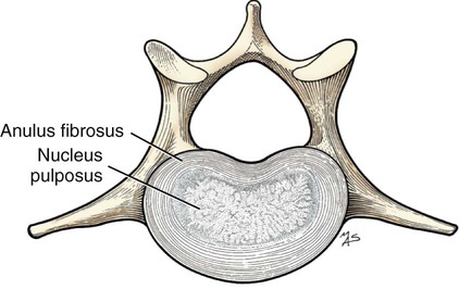
Intervertebral Discs and Short Ligaments of the Vertebral Column


The intervertebral discs (disci intervertebrales) are interposed in every intervertebral space (except between C1 and C2), uniting the bodies of the adjacent vertebrae (Figs. 5-8 and 5-9). In the sacrum of young specimens, transverse lines indicate the planes of fusion of the discs with the adjoining vertebral bodies. The thickness of the discs is greatest in the cervical and lumbar regions, the thickest ones being between the last few cervical vertebrae. The thinnest discs are in the caudal region. Those between the last few segments being smaller in every way than any of the others. Each intervertebral disc consists of an outer laminated fibrous ring, the anulus fibrosus and a central, amorphous, gelatinous center, the nucleus pulposus. The nucleus pulposus of a young dog is proportionally larger than that of an adult and more mucoid than fibroid for 1 to 7 years (King & Smith, 1955). It is a mass of mesodermal cell remnants of the notochord in a homogeneous basophilic intercellular material. Eventually small foci of degeneration and fibrosis occur, which make the disc appear opaque rather than gelatinous and may obscure the boundary with the annulus fibrosus. In chondrodystrophic breeds a chondroid degeneration may occur in young adults that eventually calcifies. There may be ossification within the disc without any adverse effect on surrounding tissues. However, the loss of function of the nucleus pulposus may result in tearing of the anulus fibrosis dorsally with protrusion or extrusion of degenerate nuclear material into the vertebral canal. This is a common cause of discomfort with or without neurologic deficits caused by spinal cord compression in these chondrodystrophic breeds. A similar but fibroid degeneration may occur in nonchondrodystrophic breeds at an older age.


< div class='tao-gold-member'>

Stay updated, free articles. Join our Telegram channel

Jul 18, 2016 | Posted by in PHARMACOLOGY, TOXICOLOGY & THERAPEUTICS | Comments Off on Arthrology

Full access? Get Clinical Tree

Get Clinical Tree app for offline access