Chapter 13 Anthrax in Free-Ranging Wildlife
Anthrax is an infectious, frequently fatal disease of domestic and wild animals and humans, caused by the gram-positive, nonmotile, endospore-forming bacterium, Bacillus anthracis. Anthrax is one of the oldest infectious diseases known to humans, and the biblical fifth and sixth plague, that first affected livestock and then humans were probably anthrax. From earliest historical records up until the development of an effective vaccine midway through the 20th century, anthrax was one of the foremost causes of uncontrolled mortality in cattle, sheep, goats, horses, and pigs worldwide.22 A sharp decline in anthrax outbreaks occurred in livestock during the 1930 to 1980 era as a result of successful national vaccination programs in many parts of the globe, as well as the subsequent advent of antibiotics. More recently, however, a resurgence of this disease in livestock has been reported in some regions, where complacency and a false sense of security have derailed vaccination programs. Some animal health regulators have become forgetful of the environmental resilience of this organism, its endemic persistence, and circulation in certain wildlife-dominated ecosystems.
The life history of B. anthracis differs markedly from most other pathogenic bacteria in that its replication and persistence appear to depend on extreme virulence and acute death of its hosts, where after it survives as a highly resistant spore during prolonged periods outside a host.3 Anthrax is an internationally reportable disease and, in 2009, the OIE (World Organization for Animal Health) reported that anthrax was still present in most countries of Africa and Asia, a number of European countries, certain countries and areas of North, Central, and South America, and certain areas of Australia. Many outbreaks of anthrax in wildlife go undetected and unreported because of surveillance inadequacies and the difficulties associated with disease monitoring in free-ranging wildlife.
Recorded Wildlife Host Range
Hugh-Jones and de Vos have listed a range of wild felids, canids, ursids, viverids, and mustelids that have been reported to have died of anthrax in zoological gardens. These deaths were most commonly associated with the inadvertent feeding of anthrax-infected carcasses, meat, or bone meal to exhibited animals. They also listed 4 free-ranging perissodactylids, 24 artiodactylids, 9 carnivore, and 2 primate species confirmed with natural infection in southern Africa.12
On a broader geographic scale, Cormack Gates and colleagues3 have also listed 23 free-ranging bovids and 6 cervid species confirmed to have died of anthrax, as well as several free-ranging zebra and rhinoceros species, Asian and African elephants, 17 free-ranging carnivores, and 2 free-ranging primate species worldwide.
Bio-Ecologic Considerations
Organism and Sources of Infection
During the interoutbreak periods, the anthrax bacterium survives in the environment as a highly resistant spore. Anthrax spores survive best in alkaline soils that are rich in calcium and have a relatively high moisture and organic content. The characteristics of dormancy and resistance to environmental factors displayed by these spores are a function of their structure, especially their hydrophobic exosporium and spore coat. The low water content of the spore confers resistance to heat and ultraviolet light.16 Calcium cations appear to participate in maintaining dormancy, as well as in the germination process. In combination with dipicolinic acid (DPA), calcium forms an extensive salt lattice that effectively immobilizes enzymes, DNA, and other metabolically active components in the core, maintaining metabolic dormancy and conferring heat resistance to core components.9
After entering the host’s body, germination appears to be triggered by moisture and warmth, as well as the presence of l-alanine in the blood serum. After germination has occurred, the vegetative bacilli undergo exponential replication, resulting in septicemia; it is the bacterial exotoxins that cause the pathology that results in the death of the animal (see later, “Pathogenesis”).
A common misconception about B. anthracis is that vegetative cells (bacilli) require the presence of atmospheric oxygen for sporulation to occur. In all Bacillus species, sporulation is a response to low nutrient conditions or dehydration, which effectively limits the diffusion of nutrients to the bacillus. The anaerobic conditions within a carcass prevent the anthrax bacilli from replicating or sporulating, even though they are in a nutrient-rich medium. In addition, putrefactive anaerobic bacteria from the gastrointestinal tract start to decompose the carcass rapidly. The vegetative form of B. anthracis are susceptible to competition from other microbes and, if putrefactive organisms reach them before the carcass is opened, they are quickly eliminated.23,25 In nature however, carcasses are rarely left intact long enough for putrefaction to eliminate all the vegetative anthrax bacilli. Instead, opening the carcass helps disperse vegetative B. anthracis into aerobic microenvironments where, either through metabolic activity or dehydration, nutrients become limited and sporulation can proceed.2 Pools of blood and tissue fluids, under aerobic conditions around the carcass site, favor sporulation. Many of these spores remain at the site of the dead animal, but some may be dispersed by water runoff, wind, and scavengers. Following sporulation, the cycle of environmental dormancy of the organism repeats itself.
Pathogenesis
It is well established that B. anthracis is not a highly invasive organism. Research results from many sources indicate that LD50 values for anthrax challenge are much higher by the oral or inhalation routes than via the parental route. However, once the anthrax spore has entered the mammalian body and germinated, it undergoes exponential replication within regional nodes, and then passes via the lymphatic vessels into the bloodstream. Bacilli that have entered the bloodstream are taken up in other parts of the reticuloendothelial system, particularly the spleen, to establish secondary centers of infection and proliferation.17 The vegetative anthrax bacilli produce a lethal combination of exotoxins, responsible for the severe clinical signs and postmortem lesions seen in anthrax. The toxin complex consists of two separate protein toxins, designated edema factor (EF) and lethal factor (LF), and a cell receptor–binding protein called protective antigen (PA).11,13 The toxin complex acts to reduce phagocytosis, increase capillary permeability, and damage blood-clotting mechanisms. The net effect produces massive edema (including the lungs and brain), hemorrhage, renal failure, and terminal hypoxia.
Routes of Infection, Transmission, and Seasonality
Anthrax outbreaks are commonly associated with low-lying depressions and rock land seep areas with high moisture content, high organic content, and an alkaline pH. Successive cycles of flood runoff and evaporation appear to concentrate the anthrax spores in these depressions, which may be referred to as concentrator areas.7 With the seasonal decrease in water levels in these concentrator areas, the resident wildlife using this water source may be increasingly exposed to higher concentrations of accumulated spores.
Transmission of anthrax relies on the ingestion of infected spores or parental inoculation of spores. Ingestion of spores is generally associated with drinking from a contaminated water source or ingesting contaminated grazing, browse, flesh, or bones; oral infection is probably the most common route of infection in wild animals. In predators and porcines, edematous lesions generally develop in the oral and pharyngeal areas, whereas in domestic and wild ruminants, necrohemorrhagic lesions develop in Peyer’s patches or segmental regions of the small intestine, eventually progressing to septicemia.14,15 Osteophagia by pregnant or lactating animals, or animals on rangeland with phosphate-deficient soil, is an important mechanism of infection in certain regions.
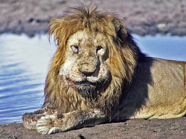
Anthrax may also penetrate broken skin or mucous membranes, and this route of infection is most commonly seen in humans who have handled anthrax-infected animal products. The animal equivalent is infection of subcutaneous tissues, generally as a result of mechanical transmission by contaminated mouthparts of biting insects. Cellulitis characterized by subcutaneous swellings is particularly common in equines. In addition, in carnivores, the massive facial and oral edema and necrosis is thought to be caused by penetration of oral or pharyngeal mucous membranes by bone spicules while chewing the bones of infected carcasses (Fig. 13-1). Following percutaneous penetration, the spores germinate and give rise to a small edematous area containing capsulated vegetative bacilli. The lesion then progresses in size, macrophages and fibrin deposits appear, lymphatics dilate, and fragmentation of connective tissue occurs, with increasing edema.4 Phagocytosis appears minimal, and the infection then progresses into a lymphangitis followed by lymphadenitis. If the infection is not halted at this stage, it may become systemic and result in fatal septicemia.
Inhalation infection is probably the least common route of infection in free-ranging wild animals living in the open air because the natural anthrax spores tend to clump together with surrounding organic material and are not easily aerosolized. However, experimental infections have demonstrated that the spores do not germinate in the airways, but are phagocytosed by mobile macrophages, which then migrate to the tracheobronchial lymph node.10 Germination begins once the infected macrophages have arrived in the lymph node, and the vegetative cells freed from the phagocytes then proliferate, causing severe lymphadenopathy and rapid septicemia.20 Inhalation anthrax generally has a very rapid fatal course.
Anthrax in North American Wildlife
Parts of southwest Texas (Val Verde, Uvalde, and Webb counties) have a historic presence of endemic anthrax. This region has shallow, lime-rich, humus soils overlying limestone, which is ideal for anthrax spore survival. This endemic state is periodically punctuated with epidemic flare-ups during hot, dry summer weather, usually following heavy spring and early summer rains. These outbreaks frequently involve white-tailed deer (Odocoelius virginianus) and unvaccinated cattle, and mortalities can be spectacular. Biting flies, locally called charbon flies, are thought to be important in propagating the epidemic by mechanical transmission of anthrax bacilli from bacteremic animals, resulting in centrifugal spread of infection.12
Anthrax in Wildlife in Sub-Saharan Africa
In the southeastern subtropical savannahs of South Africa and Zimbabwe, anthrax is a multispecies disease, with mortalities recorded in 36 wild species. However, analysis of the outbreaks in this ecosystem has shown that greater kudu (Tragelaphus strepsiceros) are the most important anthrax host, and that contamination of natural browse by blowflies (Chrysomyia and Lucilia spp.) is the most important transmission mode during these anthrax epidemics. Kudu carcasses are totally overrepresented in the carcass counts during outbreaks, which is illustrated by the fact that in the Kruger National Park, where this species constitute only 3.7% of the total large ungulate population, during four anthrax outbreaks between 1990 to 1999 they represented 42% to 62% of the anthrax-positive carcasses found.1 In addition, the fact that kudu develop very high-terminal bacteremias and have thin skins that are easily opened up by most scavengers, makes kudu an important anthrax amplifier in this ecosystem.6 Nyala (Tragelaphus angasi), another browsing antelope, are also frequently infected during outbreaks in the localities where they occur.
In the more arid southern African savannahs of Botswana and Namibia, elephants (Loxodonta africana), African buffaloes, zebras, and springbok (Antedorcas marsupialis) are important role players, and transmission appears once again to be mainly via infected water or grazing related to dry season resource stress and population clustering. The situation in Etosha National Park in northern Namibia is unique in that anthrax is endemic in this park and cases are confirmed in most years. In addition, this park has temporal clustering of cases in elephants at the end of dry season, followed by a summer (wet season) outbreak in plains ungulates such as zebra, wildebeest (Connochaetes taurinus), and springbok, probably related to contamination of grazing by carcasses or contamination of rain pools by wading vultures.8,17
Stay updated, free articles. Join our Telegram channel

Full access? Get Clinical Tree


