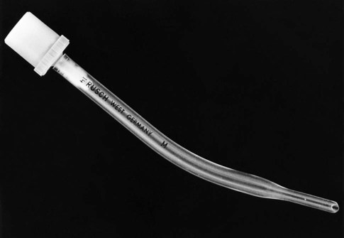
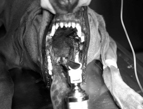
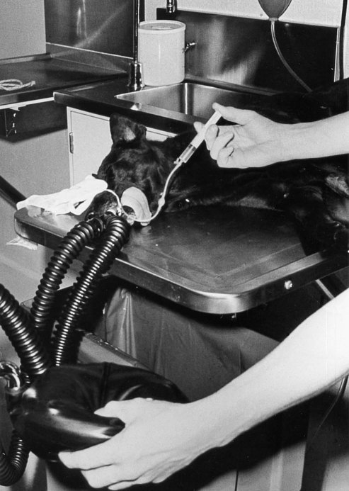
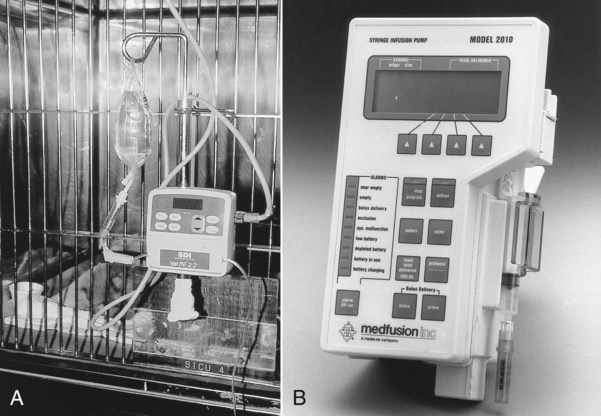
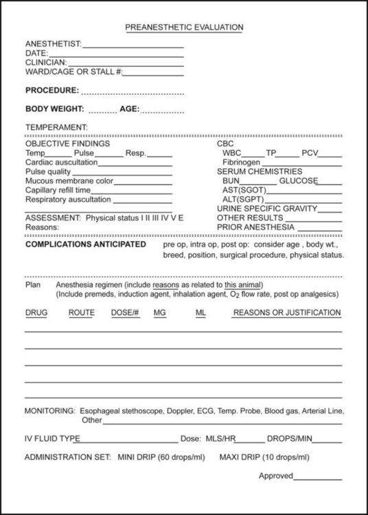
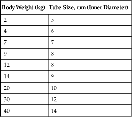
I A variety of anesthetic procedures and techniques can be used to safely produce chemical restraint and anesthesia in dogs. Local anesthetic techniques (see Chapter 7) should be considered to augment analgesia. II The choice of anesthetic regimen is influenced by the following characteristics: C Health and physical condition D Procedure (medical; surgical) E Anticipated duration of anesthesia F The familiarity of personnel with the drugs being used III Whenever possible, drugs that are reversible should be used IV Endotracheal intubation should be performed whenever possible to ensure a patent airway V Careful monitoring is mandatory to recognize and treat untoward drug effects VI Food and water should be withheld for approximately 4 to 6 hours before surgery, except in very small, very young, or diseased dogs I Review history and current drug therapy II Perform a physical examination III Review available laboratory data IV Formulate a specific anesthetic plan (Fig. 20-1) V Intravenous (IV) catheter placement is recommended VI Gather appropriate equipment and supplies B Endotracheal tube (see Chapter 11) 1. Select diameter of cuffed endotracheal tube based on the dog’s size, breed, and procedure; brachycephalic breeds usually have smaller-diameter trachea (Table 20-1); a reinforced tube (kink resistant) should be used for procedures that may require severe curvature of the neck (e.g., ophthalmic surgeries, cerebrospinal fluid tap) TABLE 20-1 Endotracheal Tube Size for Dogs 2. Check cuff for leaks by inflating with air; deflate before intubation 3. Use a stylet for small-diameter, endotracheal tubes, and reinforced tubes 4. Use an uncuffed endotracheal tube in very small dogs (Fig. 20-2) 1. Aids intubation by allowing visualization, illumination, and manipulation of tongue and laryngeal structures D Anesthetic machine and breathing system 1. Size and type of system determined by dog’s size 2. Rebreathing bag should be approximately five times the tidal volume. Tidal volume (VT) is 10 to 15 mL/kg of body weight (e.g., 5 kg × 10 mL/kg = 50 mL VT) 3. Refill carbon dioxide absorbent canister if material is exhausted (discolored or dry) 4. Evaluate anesthetic system for possible malfunctions (see Chapter 12) a. Fill vaporizer and check operation b. Turn on flowmeters and check for free movement of indicator balls or slides c. Close pressure relief valve and pressurize the system using oxygen flush valve to 20 to 30 cm water and maintain for 15 seconds; check for leaks (Fig. 20-4) 5. Connect the waste gas scavenging system to the anesthetic circle or nonrebreathing system a. Connect to “house” oxygen system, if present b. Attach smaller (E) tanks to machine; change oxygen tank if pressure gauge reads less than 500 pounds per square inch (psi) 2. Nitrous oxide (used to produce analgesia) 1. IV catheter (16- to 23-gauge) 3. Fluid administration set (standard set 10 drops/mL, pediatric 60 drops/mL). Use 60 drop/mL on dogs less than 5 kg. Administer at 5 to 10 mL/kg/hr. 4. Fluid infusion pump (Fig. 20-5) H Monitoring equipment (see Chapter 14) 2. Doppler with sphygmomanometer or oscillometric blood pressure monitor 6. End-tidal gas monitoring (oxygen, carbon dioxide, inhalant)
Anesthetic Procedures and Techniques in Dogs
Overview
General Considerations
Preanesthetic Evaluations (See Chapter 2)
Body Weight (kg)
Tube Size, mm (Inner Diameter)
2
5
4
6
7
7
9
8
12
8
14
9
20
10
30
12
40
14
![]()
Stay updated, free articles. Join our Telegram channel

Full access? Get Clinical Tree


