Anemia

Chapter 120 Anemia


KEY POINTS


SIGNALMENT AND HISTORY


The breed of the patient may be of particular importance now that many hereditary blood diseases and genetic predispositions in certain breeds have been recognized (e.g., immune-mediated hemolytic anemia [IMHA] in Cocker Spaniels). Although many hereditary erythrocyte defects,2,3 coagulopathies (e.g., hemophilia A and B, factor VII or XI deficiency), von Willebrand disease, and thrombopathias lead to anemia in juvenile animals, some may be recognized only in the older animal after acute, and possibly recurrent, episodes of bleeding have occurred.2,4,5 Furthermore, most hereditary disorders are observed in equal proportions in both genders, although hemophilia A and B, two serious coagulopathies, affect only male animals (X-chromosomal recessive trait).


A carefully taken history and review of previous laboratory test results can often provide clues as to the duration and cause of the anemia. For instance, depending on the geographic location and travel history, exposure to a certain infectious agent such as Babesia, Ehrlichia, Leishmania, and Leptospira spp in dogs and various viral (feline leukemia virus, feline immunodeficiency virus, and feline infectious peritonitis) and bacterial (Mycoplasma haemofelis, other Mycoplasma spp) and parasitic (Cytauxzoon felis) infections in cats (as well as other emerging infectious diseases) may require diagnostic testing by serology, antigen assay, or polymerase chain reaction test. There are heavy metals (zinc and copper) and other chemicals (anticoagulant rodenticides), drugs (e.g., antithyroid drugs [cat], estrogens [dog], heparin, warfarin, aspirin), and even food components (onions, garlic) that represent known triggers of anemia (see Intoxications section). A history of anemia, hemorrhage, icterus, or requirement for transfusion therapy may indicate a recurrent problem. Finally, some concurrent chronic illnesses and organ disorders, such as renal and hepatic failure, diabetes mellitus, or adverse effects of medical therapy (e.g., chemotherapeutics and many other drugs, hypophosphatemia induced by insulin administration, or hyperalimentation) may lead to severe anemia.


CLINICAL SIGNS


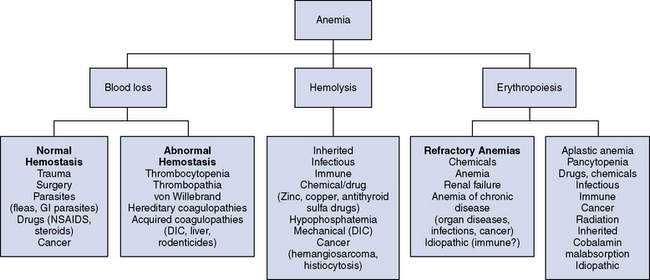
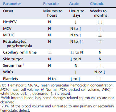
The clinical signs of anemia vary greatly depending on the rapidity of onset, type, and underlying cause. It is of utmost importance to determine if hemorrhage, hemolysis, or a hematopoietic production disorder is causing the anemia. Any form of anemia may be associated with pallor, and this may be the only sign in some animals. However, characteristic signs such as hemorrhage with blood loss–induced anemia (Table 120-1) or icterus or pigmenturia with hemolytic anemia may help the clinician determine the etiology.


In animals with peracute blood loss, the clinical signs are mostly related to hemorrhage and hypovolemia (hypovolemic shock), and animals with acute and chronic anemias display the more typical signs (e.g., lethargy, pallor, tachycardia, tachypnea). External blood loss is often readily evident, except for animals with gastrointestinal (GI) hemorrhage originating from the nasopharynx to the rectum. In contrast, internal hemorrhage may be hard to localize, especially in animals with retroperitoneal or deep muscle hemorrhage. A single site of hemorrhage may occur secondary to a local process such as trauma or surgery (e.g., a single lacerated vessel, hematomas), but may also be caused by an underlying bleeding tendency. Surface hemorrhage, such as petechiations and ecchymoses, suggest a platelet disorder (thrombopathia or thrombocytopenia), whereas multiple or recurrent hematomas and intracavitary hemorrhage indicate a coagulopathy. von Willebrand disease and hereditary coagulopathies occur commonly in dogs.


Icterus may be observed the day after substantial hemolysis and the serum bilirubin concentration often exceeds 2 mg/dl. However, pigmenturia due to hyperbilirubinuria, and less commonly hemoglobinuria, is noted earlier in the disease process, even with mild hemolytic disease. It should be noted that animals with IMHA may have a combination of cholangiohepatic and hemolytic processes that result in severe icterus. Finally, cyanosis is not a clinical sign of anemia except in the presence of methemoglobinemia associated with oxidative injury to the RBCs, such as acetaminophen toxicity in cats. There must be approximately 5 g/dl of unoxygenated Hb in the capillaries for the blue color of cyanosis to be appreciated clinically; therefore severely anemic animals with severe hypoxemia may not appear cyanotic despite severe concurrent hypoxemia when having concurrent cardiopulmonary disease. The tissue hypoxia caused by anemia also activates a series of compensatory mechanisms that result in the typical signs of anemia and include1:


Stay updated, free articles. Join our Telegram channel

Sep 10, 2016 | Posted by in SMALL ANIMAL | Comments Off on Anemia

Full access? Get Clinical Tree

Get Clinical Tree app for offline access