CHAPTER 26Anatomy and Examination of the Normal Testicle
THE SCROTUM
The testes of the normal stallion are contained within the slightly pendulous scrotum located between the stallion’s hindlimbs. In a normal stallion, the scrotum and contents should appear generally symmetric. The scrotum is formed from an outpouching of skin, with two distinct pouches separated by a median raphe. The scrotum contains the two testes, epididymides, spermatic cords, and cremaster muscles. The wall of the scrotum consists of four layers: (1) skin, (2) tunica dartos, (3) scrotal fascia, and (4) tunica vaginalis. The scrotal skin is essentially hairless and contains large numbers of sebaceous and sweat glands, which assist with thermoregulation of the testicles.1–3 A longitudinal scrotal raphe marks the center line of the scrotum and continues cranially onto the prepuce and dorsocaudally onto the perineum. The tunica dartos layer consists of both muscular and fibroelastic tissue and is adhered to the inner surface of scrotal skin, lining both scrotal pouches and reflecting along the raphe to form the median septum. The degree of relaxation or contraction of this layer results in alterations in shape and positioning of the scrotum in relation to the body wall, assisting with testis thermoregulation.3 The scrotal fascia is a loose connective tissue layer between the tunica dartos and tunica vaginalis and allows the testis to move freely within the scrotal pouch.3 The tunica vaginalis is the innermost layer of the scrotum and is formed by evagination of the peritoneum during testicular descent. This layer is closely apposed to the tunica albuginea of the testis, but separated by a potential space termed the vaginal cavity. A thin layer of fluid lines the vaginal cavity to permit the testis to slide smoothly within the scrotum. On palpation the scrotal skin should be thin, consistent throughout without wrinkling, slide loosely over the testicles and be free of any lesions. The tunics should palpate as smooth layers without wrinkling.
THE TESTIS
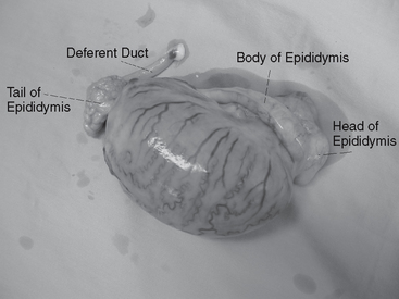
The testis is encapsulated by the tunica albuginea, a layer of collagenous tissue and smooth muscle (Figure 26-1). Trabeculae of the tunica albuginea extend into the testicular parenchyma to provide a supporting framework and divide the testis into lobules.3 The mediastinum testis of the stallion is located at the cranial pole of the testis where the excurrent ducts cross the tunica albuginea and enter the head of the epididymis. This results in a mediastinum testis that is less obvious ultrasonographically compared with other species such as the bull, where the mediastinum testis is oriented axially within the testis. On manual palpation the testes of a normal stallion lie horizontally within the scrotum with the tail of the epididymis directed caudad. Palpation of the tail of the epididymis and the ligament of the tail of the epididymis assist the examiner in determining normal orientation of the testicle. The ligament is palpable as a fibrous nodule attaching the tail of the epididymis to the caudal pole of the testis. The tone of the testicles on palpation should be firm to rigid and resilient. Excessive softness or firmness may be indicative of disease conditions.
TESTIS SIZE AND VOLUME
Testicular size varies among stallions depending upon breed, size, age, season, and reproductive status. Each testis of a normal postpubertal light horse breed stallion weighs between 150 and 300 g.4 Testis size and parenchymal weight correlate highly with daily sperm production and is a useful predictor of a stallion’s breeding potential. Because parenchymal weight cannot be measured in the live stallion, testicular measurements are used. Individual testis measurements have been shown to correlate well with parenchymal weight (r2= 0.57 to 0.82) and daily sperm output (r2= 0.52 to 0.76).5–8 Testicular measurements therefore become a critical component of a breeding soundness evaluation in the stallion. The testes are measured individually using calipers for length, width, and height. Alternatively, ultrasonographic measurements can be taken to determine cross-sectional area (through the widest portion of the testicle) and length. In addition, the total scrotal width is measured using calipers. The testicles should be pushed downwards into the scrotum with one hand while the other hand operates the calipers. The average of three measurements should be used to reduce operator error. Recommended minimum guidelines for light horse stallions9 for total scrotal width and individual testicular measures are given in Tables 26-1 and 26-2, respectively. The reproductive characteristics, including testicular measurements, of miniature stallions have also been determined.10Tables 26-3 and 26-4
Stay updated, free articles. Join our Telegram channel

Full access? Get Clinical Tree


