25 Alteration in pupil function
INTRODUCTION
These two cases illustrate altered pupil function localized to different regions of the nervous system.
CASE HISTORY
The dog suddenly became mentally dull and ataxic 5 days prior to referral. The dog bumped into objects, growled at the owner, slept a lot more and urinated indoors. Appetite and thirst had decreased. The dog fell to the left when getting off the sofa.
CLINICAL EXAMINATION
The dog was obtunded with a lowered head posture. The left menace response was absent and the pupils were bilaterally mydriatic and lacked a PLR. Fundic examination was normal. The dog walked slowly and on one occasion was seen to lean to the left. Hopping was slightly slower in all limbs. Proprioception was normal.
NEUROANATOMICAL LOCALIZATION
The lesion was localized to the midbrain to explain the dilated pupils lacking a PLR. An absent menace can be due to a depressed mental state, however the change should be bilateral. A left cerebellar lesion could explain the left menace deficit and falling to the left. A right cerebral lesion or left optic nerve lesion could also account for the menace deficit in this dog in which case the lesion may be in the region of the optic chiasm and compressing the thalamus.
CASE WORK-UP
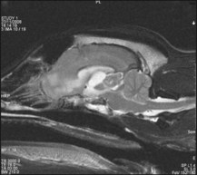
MRI found a contrast-enhancing lesion which was isointense on T1 and of variable intensity on T2WI. There was secondary dilation of the third and lateral ventricles (Fig. 25.1). Within the third ventricle choroid plexus papilloma (CPP) and ependymoma are intraventricular in location and enhance uniformly. Meningiomas can be intraaxial.
CASE HISTORY
The dog had been lethargic for 1 week, lagging behind on exercise and lacking interest on a walk. He had vomited twice. Two days prior to referral, bilateral protrusion of the third eyelids occurred, followed the next day by squinting in the light and bilateral dry nasal discharge. The dog appeared to be urinating more often but was not polydipsic.
CLINICAL EXAMINATION
The dog had a depressed mental state, dry oral mucous membranes and bilateral dry nasal discharge. There were normal menace responses and absent PLRs bilaterally. The pupils were midsized with the right slightly smaller than the left. The third eyelids were prolapsed. Right enophthalmos and right ptosis were present.
Gait, posture, proprioception, hopping and spinal reflexes were all normal.
The Schirmer Tear Test (STT) was 0 mm bilaterally.
A 10% phenylephrine testing of the right eye produced a normal palpebral fissure size and third eyelid appearance in 5 minutes.
NEUROANATOMICAL LOCALIZATION
Dry mouth (xerostomia)
Lack of saliva impedes the formation of a food bolus but xerostomia is not a reported cause of dysphagia in small animals. A unilateral facial nerve lesion commonly results in dry eye but it would be impossible to clinically recognize decreased saliva production from the same lesion.
Dry nose (xeromycteria)
Dry nose presents as a dry nasal plane (unilateral or bilateral), epithelial crusts and a slight mucopurulent discharge.
Serous glands of the nasal plane have a parasympathetic innervation by the facial nerve. The afferent stimulus for nasal secretion is transmitted by the trigeminal nerve. Pathological dryness of the nose, xeromycteria, results from:

Full access? Get Clinical Tree

