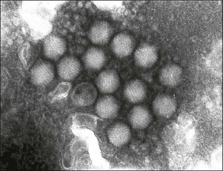
Chapter 48 Adenoviruses (from the Greek word adenos meaning gland) were first isolated from explant cultures of human adenoids. They are non-enveloped, icosahedral viruses, 70–90 nm in diameter, containing a single linear molecule of double-stranded DNA (Fig. 48.1). One to two protein fibres protrude from each of the 12 vertices of the capsid. The family Adenoviridae contains five genera designated Mastadenovirus, Aviadenovirus, Atadenovirus, Siadenovirus and a genus of fish adenoviruses, Ichtadenovirus. Mammalian adenoviruses, assigned to the genus Mastadenovirus, share a common antigen and are serologically distinct from those that infect birds, genus Aviadenovirus. Many adenoviruses agglutinate rat or monkey erythrocytes. The fibre protein is responsible for this property and contains type-specific determinants. The haemagglutination inhibition test is used to confirm serospecificity. Adenoviruses are moderately resistant, surviving in the environment for days or weeks under suitable conditions. Adenoviruses can withstand freezing, mild acids and lipid solvents. Infectivity is lost following heating at 56°C for more than 10 minutes. The adenoviruses of veterinary importance are shown in Figure 48.2 and Table 48.1. Table 48.1
Adenoviridae
Virus
Host species
Disease
Canine adenovirus serotype 1
Dog, fox
Cause of infectious canine hepatitis, a generalized infection affecting liver and blood vascular system
Canine adenovirus serotype 2
Dog
One of the causes of infectious tracheobronchitis (kennel cough), a highly contagious respiratory disease complex
Equine adenovirus A
Horse
Generally subclinical or mild respiratory infection but also associated with fatal pneumonia in Arabian foals affected by combined immunodeficiency disease
Bovine adenoviruses
Cattle
Occasional outbreaks of enteric and respiratory disease
Ovine adenoviruses
Sheep
Occasional outbreaks of enteric and respiratory disease
Porcine adenoviruses
Pig
Occasional cases of diarrhoea, usually subclinical
Fowl adenoviruses
Fowl, quail
Numerous serotypes. Frequently isolated from healthy birds or following mild respiratory disease. Associated with inclusion body hepatitis and quail bronchitis
Duck adenovirus A
Fowl, duck
Cause of egg drop syndrome in laying hens
Turkey adenovirus A
Turkey, pheasant
Cause of turkey haemorrhagic enteritis which affects four- to 12-week-old turkey poults and of marble spleen disease which affects two- to eight-month-old pheasants
![]()
Stay updated, free articles. Join our Telegram channel


Full access? Get Clinical Tree


