Abdominal cavity

Chapter 4


Abdominal cavity


4.1 Hernias


Definitions


A hernia comprises the protrusion of an organ or part of an organ through the wall of the cavity normally containing it. Hernias may be classified as direct or indirect, internal or external.


A direct hernia occurs through a rent or tear whereas an indirect hernia occurs through a natural passage such as the inguinal canal or umbilicus.


An external hernia occurs through the body wall producing a visible and palpable swelling covered by skin, whereas an internal hernia occurs within the abdominal cavity (Box 4.1).


Umbilical hernias


Umbilical hernias are significantly more common in fillies than in colts, and may be hereditary. Because the majority of small hernias eventually close spontaneously it has become customary to wait until the foal is 6–12 months old before attempting surgery. Large defects show little tendency to spontaneous closure, and early repair is rewarded by the greater ease with which repair can be achieved in the young foal.


Congenital umbilical hernias are present at birth and are due to failure of the abdominal wall to close. Acquired umbilical hernias develop at 3–4 weeks of age. Excessive straining to defecate or micturate, and infection of the umbilicus, may be causative factors. It has been suggested that umbilical hernias are often the result of improper management/handling of the foal at birth such as manual ligation of the cord.


The fibrous hernial ring is composed of the aponeurosis of the transverse muscle, the fused oblique muscles and the abdominal tunic.


Umbilical hernias may be oval in shape and vary in size from one to several centimetres in diameter.


The hernial sac comprises an inner peritoneal layer and an outer layer of skin linked by varying amounts of connective tissue. Intestine (small intestine or caecum), omentum or both may constitute the contents of the hernial sac.


Incarceration and strangulation of the herniated intestine is rare and most likely to occur when the hernial ring is intermediate in size (i.e., large enough to allow entry of intestine yet small enough that intestinal distension results in constriction of blood vessels and lymphatics).


A parietal or Richter’s hernia is defined as incarceration of the antimesenteric portion of intestinal wall is trapped within a hernial orifice. The lumen of the intestine bowel is not completely obstructed, and pain may be less severe. If the hernia goes unrecognized and uncorrected, ischaemic necrosis of the incarcerated ‘pouch’ of intestinal wall may lead to the development of an enterocutaneous fistula. The ileum and less frequently caecum may be involved.


Umbilical hernias complicated by infection of the remnants of the umbilical stalk are encountered only rarely in foals compared with calves. The umbilical structures should close within a week of birth and only fibrous remnants should remain at 6 weeks. In the event of infection one or more of the umbilical structures (one umbilical vein, two umbilical arteries and the urachus) will remain patent. Ultrasonography provides the best means of identifying which structure(s) is involved.


Examination of the hernia


In the vast majority of cases, gentle pressure on the protruding swelling will reduce the hernia, allowing identification of the hernial ring, which should be assessed for size, shape and rigidity.


Incarcerated hernias present as tense, painful swellings which are usually irreducible. Occasionally gas in the entrapped intestine may have been dispersed during transport to a surgical facility or by gentle pressure allowing reduction to be achieved. Although the immediate risk to the foal’s life may have been removed, whenever possible repair of the hernia should be carried out as soon as possible. Immediate repair will allow identification and removal of necrotic intestine which will eventually rupture.


Incision of the hernia sac and careful examination of the caecum and small intestine should be performed to rule out the possibility of ischaemic compromise which is particularly likely with a Richter hernia. Even if ischaemic damage is not considered likely, any delay in repair of the hernia should be kept to a minimum because of the possibility of a further episode of incarceration which on that occasion may progress to strangulation.


Closed reduction


A wide variety of methods of treatment have been used in the past, including blisters, transfixing skewers and wooden or metal clamps. Although clamps are still in use, the method of closed reduction most often used is the application of elastrator rings at the base of the sac after ensuring that any contents have been returned to the abdomen. There may be some oedema between the rubber ring and the abdominal wall on the day after application, but any local inflammation usually resolves in 7–10 days. Separation of the hernial sac vestige should be allowed to occur without assistance and takes approximately 3–4 weeks. This technique depends on subcutaneous fibrous scar tissue to obliterate the defect, and is only useful in treating small one-finger defects.


With the availability of safe anaesthetic techniques and improved suture materials, there is little indication for employing a method which is less than certain to correct the hernia. Open reduction, whereby the defect in the abdominal wall is closed by apposing the edges of the ring or by the use of an inlay prosthesis is a much more satisfactory method of treatment.


Open reduction


The surgery is performed under general anaesthesia and strict aseptic conditions with the patient in dorsal recumbency. An elliptical skin incision is made around the umbilical scar extending 2 cm beyond the margin of the hernial ring cranially and caudally. Care must be taken to avoid removing too much skin. The peritoneal sac is carefully dissected away from the overlying skin and the abdominal wall until the margin of the ring can be identified. Unless there is residual umbilical stalk present, or ischaemic damage to the intestine is suspected, the hernial sac need not be incised but rather simply returned to the abdominal cavity.


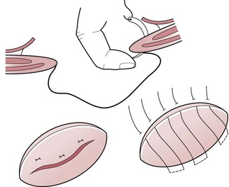
The majority of hernial rings are oval in shape and their edges are sufficiently pliable to allow them to be closed by a series of overlapping mattress (Mayo) sutures of absorbable or non-absorbable material.


The sutures are inserted 1–2 cm from the margin of the ring and are carried through the full thickness of the abdominal wall on both sides of the ring. A finger inserted into the inverted sac serves to guide the needle and prevent inadvertent damage to the intestine (Figure 4.1). The ends of each suture are held with haemostats until they are all in place. Simultaneous traction on the sutures overlap the edges of the ring and this is maintained while each individual suture is tied. The subcutaneous tissue is apposed with a continuous suture of polyglactin, and the skin with a subcuticular suture of the same material. The application of an elastic bandage encircling the abdomen (or a stent bandage in the case of small hernias) will provide protection from contamination and eliminate dead space.


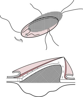
With large hernial rings, it may be necessary to bridge the defect with a prosthetic mesh or gauze. The mesh, which must be inert and nonreactive to the tissues, stimulates fibroplasia, resulting in it being enveloped in a thick fibrous bed that fills the defect and strengthens the abdominal wall.


The mesh is best placed in an extraperitoneal position between the internal rectus abdominal sheath and the peritoneum. The peritoneal sac is dissected down to the hernial ring and inverted into the abdominal cavity as described above. If the sac is sufficiently large, the mesh can be placed within it (Figure 4.2), but if it is not, the peritoneum is reflected peripherally from the deep fascial sheath of the rectus abdominis muscle for 1–3 cm to create a space for the mesh. The mesh is cut so that it overlaps the margin of the ring by the same amount. The sutures are preplaced before tying. Sufficient tension is applied so that the mesh is kept taut and flat. The subcutaneous tissues and skin are then carefully apposed over the mesh.


Ventral hernias


A ventral hernia is one that occurs through any part of the abdominal wall other than the umbilicus or inguinal canal. They may be traumatic in origin or incisional. Kicks, collisions with blunt objects and straddling gates are common causes. Common sites are low in the flank (just dorsal to the edge of the rectus muscle), along the costal arch and along the ventral abdomen.


The abdominal wall is weakest just dorsal to the rectus abdominis muscle, since the oblique abdominal muscle is aponeurotic here, and only the transverse abdominal muscle is present. Flank trauma usually causes the abdominal wall to split just above the rectus muscle. Usually the tear in each layer occurs along the direction of the fibres, and if damage to the external oblique is extensive, the inguinal ring may be involved. If the peritoneum is also torn, herniated intestine will come to lie beneath the skin and panniculus muscle.


A large tear will allow a large mass of viscera (e.g. large colon) to escape from the abdomen, but the size of the swelling is not necessarily an indication of the size of the defect because a considerable length of small intestine can escape through a small rent.


Inflammatory exudate accumulates in the surrounding tissues, but little change may occur in the herniated intestine during the first 2–3 weeks after injury. Nevertheless, the risk of strangulation of intestine necessitates early diagnosis and surgical treatment.


Diagnosis: Signs of depression, abdominal discomfort and cessation of defecation indicate intestinal obstruction requiring prompt intervention.


Diagnosis is not always straightforward, and a number of diagnostic procedures should be considered to differentiate a hernia from a haematoma or abscess:


Palpation is complicated by the associated haemorrhage and oedema. Most defects are too ventral to be palpated per rectum. Aspiration, although helpful in identifying abscesses, is not advisable because of the risk of penetrating the bowel or introducing infection into a haematoma. Auscultation may reveal intestinal sounds which are more easily heard than over the remainder of the abdominal wall. Ultrasonography provides the most useful way of detecting herniated intestine.


Treatment: When intestinal obstruction necessitates immediate surgery, the tissues surrounding the rent or hernia are very friable and have little suture holding power. It is not uncommon for disruption of the surgical repair to occur during recovery from anaesthesia. Therefore, when the hernia is not accompanied by signs of intestinal obstruction, it is advisable to delay surgery for 3–6 weeks until swelling has subsided and deposition of collagen has increased the tensile strength of the damaged tissues.


Under general anaesthesia, the skin and subcutaneous muscle are carefully excised to reveal the herniated intestine. If examination of the intestine reveals no irreversible damage it is returned to the abdominal cavity. A midline laparotomy incision is helpful in identifying the precise site of the tear and allowing traction to be applied to the herniated gut.


The defect is closed by suturing each layer in turn.


When the defect is very large or its edges are too rigid to appose by suture, satisfactory repair may be achieved using mesh. Following closure of the peritoneum and transverse muscle by suture, a piece of polypropylene mesh is sutured to the deep face of the internal oblique aponeurosis.


Incisional hernias


Incisional or postoperative hernias are encountered relatively frequently following abdominal surgery, particularly in large, heavy horses.


Improper closure of the incision, sutures breaking or tearing through tissues and, of greatest importance, postoperative wound infection, are contributory factors.


The weakened abdominal wall undergoes loss of continuity and a hernia develops often after a delay of several weeks or months. The hernial rings vary considerably in size and can be very large.


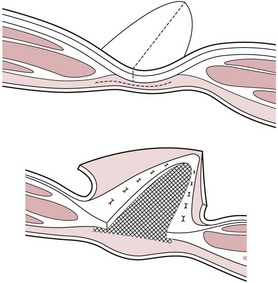
Forceful approximation of the edges of a large fascial defect inevitably leads to failure. To repair large hernias a mesh prosthesis is required. Since the margin of the defect is less well defined than in umbilical hernias, it is necessary to dissect the peritoneum from the inner sheath of the rectus to create an adequate ‘shelf’ to support the mesh (Figure 4.3). Alternatively, a mesh inlay graft with the onlay apposition of supportive hernial sac fascia can be used (Figure 4.4).


Stay updated, free articles. Join our Telegram channel

Jun 18, 2016 | Posted by in EQUINE MEDICINE | Comments Off on Abdominal cavity

Full access? Get Clinical Tree

Get Clinical Tree app for offline access