A
5-Fluorouracil Toxicosis
BASIC INFORMATION 
DIAGNOSIS 
DIFFERENTIAL DIAGNOSIS
Diseases or intoxications that could cause vomiting (see p. 1173) and/or seizures (see pp. 1009 and 1425)
INITIAL DATABASE
• CBC with differential (baseline, monitor q 24-72 hours for 20 days); initial leukocytosis followed by evidence of myelosuppression, leukopenia, pancytopenia in 5-20 days. Hematocrit increased due to dehydration then decreased due to gastrointestinal (GI) bleeding.
TREATMENT 
ACUTE GENERAL TREATMENT
• Decontamination of asymptomatic patient
○ Emesis using activated charcoal with a cathartic if patient is showing no clinical signs and the ingestion is recent (<1 hour)
• Manage tremors and seizures (see p. 1009)
○ Seizures are rarely controlled with diazepam alone (dogs: 2-5 mg/kg IV; cats: 0.5-1 mg/kg IV). If seizures persist after diazepam therapy, give phenobarbital IV bolus, 2-5 mg/kg (can be repeated at 20-minute intervals up to two times). May then add phenobarbital IV infusion (2-10 mg/hour IV, titrated to effect) to diazepam; or
CHRONIC TREATMENT
Intensive supportive care may be needed for days/weeks, especially if myelosuppression occurs.
PEARLS & CONSIDERATIONS 
COMMENTS
• Severe signs or death can result from 5-FU toxicosis; early decontamination and immediate, intensive treatment are essential, and owners should be advised of the seriousness of exposure to 5-FU.
Abdominal Compartment Syndrome
BASIC INFORMATION 
EPIDEMIOLOGY
RISK FACTORS
• Any patient with abdominal disease or accumulations of fluid, gas, or tissue in the abdominal cavity can develop elevated IAP.
CLINICAL PRESENTATION
DISEASE FORMS/SUBTYPES
• Secondary ACS: extraabdominal cause, most commonly sepsis or burn patients that require aggressive fluid resuscitation
ETIOLOGY AND PATHOPHYSIOLOGY
• ACS (sustained IAP > 20 mm Hg with new organ dysfunction/failure) develops if IAH is not recognized and treated.
• ACS is not a disease, but a distinct syndrome of clinical signs and abnormalities that can have multiple causes.
• At low intraabdominal volumes, the abdominal wall is very compliant. Large increases in abdominal volume result in minor increases in IAP. At higher abdominal volumes, abdominal wall compliance decreases, and even small increases in volume can cause major increases in IAP.
• Increased pressure in the abdomen leads to hypoperfusion and ischemia of abdominal organs and, when severe, direct compression of blood vessels. This leads to alterations in renal (most recognized), hepatic, and gastrointestinal functions. Anuria can occur with IAP > 30 mm Hg. Liver function, including cytochrome P450 function, is impaired. Intestinal edema can develop, as well as bacterial translocation through gastrointestinal walls.
• Reduced ability for the diaphragm to contract against the tense abdomen impairs tidal volume and compromises ventilation, resulting in hypoxia and hypercapnia. Mechanical/positive pressure ventilation is similarly affected.
• Increase in central venous pressure (CVP) and drop in cardiac output (increased systemic vascular resistance, decreased venous return to the heart, compression of vascular structures from increased intrathoracic pressure) result in altered cardiovascular function.
• Hormonal changes occur in response to the altered cardiovascular state, including elevations in renin-angiotensin activity, antidiuretic hormone, and catecholamine levels.
• Ischemia-reperfusion injury occurs after shock resuscitation and can affect distant organ function.
• Abdominal wall complications can occur secondary to hypoperfusion, including delayed or impaired wound healing, dehiscence, and surgical site infection.
• Recognition of increasing IAP and prevention of ACS are crucial to reducing morbidity and mortality.
DIAGNOSIS 
DIAGNOSTIC OVERVIEW
IAP must be measured to confirm the diagnosis of abdominal compartment syndrome.
INITIAL DATABASE
• A water manometer is attached to a stopcock in the evacuation line from a standard urinary (urethral) catheter.
• The urinary bladder is emptied, and a few milliliters of isotonic sterile saline are placed in the bladder (3-10 mL, depending on patient size).
• After 30-60 seconds (to allow the saline to warm and the detrusor muscle to relax, avoiding a falsely elevated reading), the zero mark on the manometer is held at the level of the cardiac atria (level of the umbilicus in lateral recumbency).
• Abdominal muscle contractions can increase IAP readings. Obese patients may have a higher baseline IAP.
• Alternative measurement techniques include nasogastric balloon catheter (good for continuous measurement), direct intraabdominal catheter/abdominal drain, and venous catheter in the caudal vena cava. These techniques give similar information but can be more cumbersome to place and maintain.
• Trends in pressure parameters may be more important than any single number. It is important to use the same positioning each time a measurement is taken.
• The IAP measured at one point in the abdominal cavity can be assumed to be the pressure throughout the abdominal cavity.
ADVANCED OR CONFIRMATORY TESTING
• Lactate levels, liver enzyme changes, urine output, blood urea nitrogen, and creatinine levels to identify abdominal organ dysfunction associated with ACS
• Measurements of cardiac output and other cardiovascular parameters and monitoring mentation and intracranial pressure give a more extensive assessment of cardiovascular function and perfusion.
TREATMENT 
ACUTE, GENERAL TREATMENT
• Medical Management
○ Sedation, analgesia, and/or neuromuscular blockade to reduce muscle tone secondary to pain, agitation, and ventilator dyssynchrony, which result in increased IAP
PEARLS & CONSIDERATIONS 
COMMENTS
• Most critically ill patients should have urethral catheters in place to monitor urine output, which allows for IAP monitoring and early detection of ACS.
• IAH may falsely elevate indirect intracardiac pressure readings due to abdominothoracic transmission of pressure.
Abdominal Distention
BASIC INFORMATION
CLINICAL PRESENTATION
HISTORY, CHIEF COMPLAINT
• Abdominal enlargement (rate of distention is extremely variable, varies from acute to chronic and insidious)
• When ascites is present and the fluid accumulation is rapid, clinical signs of weakness and dyspnea are more likely to be apparent.
ETIOLOGY AND PATHOPHYSIOLOGY
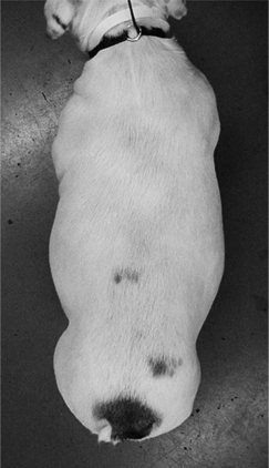
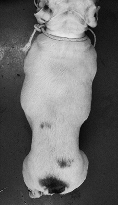
 ABDOMINAL DISTENTION Dorsal view of a young English bulldog with marked ascites caused by right-sided congestive heart failure due to severe pulmonic stenosis. The abdominal distention hides the dog’s weight loss and may be mistaken by the owner as an increase in lean body mass. This degree of abdominal distention was associated with poor appetite, lethargy, and dyspnea in this dog.
DIAGNOSIS
INITIAL DATABASE
• Abdominal radiographs
○ Mass lesions, fat, or organomegaly may be apparent or suggested by displacement of gas-filled organs.
• Thoracic radiographs
○ Enlarged cardiac silhouette and/or caudal vena cava if congestive heart failure or pericardial effusion (rule out as cause of ascites)
• CBC, biochemistry panel, and urinalysis are indicated for any animal with ascites or abnormal abdominal distention. Abnormalities depend on underlying cause and extent of organ damage.
• Abdominal paracentesis (see p. 1194) if ascites; obtain fluid for cytology and biochemical evaluation.
ADVANCED OR CONFIRMATORY TESTING
• Fine-needle aspirate or needle biopsy (mass, enlarged organs, etc.). Biopsy not recommended if area is infected (abscess, pyometra) or likely to hemorrhage (e.g., suspected hemangiosarcoma).
• Prothrombin time if anticoagulant intoxication is suspected as cause of ascites (paracentesis contraindicated if coagulopathy is suspected)
TREATMENT
TREATMENT OVERVIEW
Treatment of underlying disease and therapeutic abdominal drainage if large-volume ascites is causing the abdominal compartment syndrome (see p. 4)
PEARLS & CONSIDERATIONS
Aberrant Adrenocortical Disease (Increased Adrenal Sex Hormone Production) ![]()
BASIC INFORMATION 
DIAGNOSIS 
ADVANCED OR CONFIRMATORY TESTING
• Adrenocorticotropic hormone (ACTH) stimulation test with sex hormone panel (progesterone, 17-hydroxyprogesterone, estradiol, androstenedione, testosterone):
○ Substantial overlap in sex hormone serum concentrations between healthy, nonadrenally ill, and hyperadrenocorticism patients make sex hormone assays ineffective screening tests for hypercortisolism.
TREATMENT 
CHRONIC TREATMENT
• The following recommended by some, with anecdotally supported results (see http://www.vet.utk.edu/diagnostic/endocrinology/pdf/TreatmentInfoAtypicalCushingsRevised201001.pdf):
○ Melatonin (3-6 mg/dog PO q 12 h, based on body weight); inhibits aromatase and 21-hydroxylase enzymes
○ Melatonin implants (www.melatek.net) 8, 12, 18 mg for dogs weighing <25, 25-50, and >50 lb, respectively
POSSIBLE COMPLICATIONS
See Hyperadrenocorticism, p. 548; Adrenal Neoplasia (Adenoma/Carcinoma), p. 43
Stay updated, free articles. Join our Telegram channel

Full access? Get Clinical Tree


