CHAPTER 45. Respiratory Disorders
Rebecca S. McConnico
RESPIRATORY EXAMINATION
I. History
A. Exact complaint
1. Description of problem
2. Frequency
3. Duration
B. Type of housing and bedding
C. Are other animals sick?
D. Age of horse
1. Infections more common in younger horses
2. Chronic airway disease more common in older horses
E. Additional questions to ask
1. Associated with work?
2. Weight loss or gain?
3. Duration of ownership?
4. Recent change of environment?
5. Acute or gradual onset?
6. Previous surgery?
7. Vaccination status?
F. Observation of environment
II. Physical examination
A. Attitude
1. Depressed: Bacterial or viral infection (e.g., bacterial pneumonia)
2. Bright, alert, responsive: Allergy (e.g., summer pasture associated recurrent airway disease [RAD], such as indoor RAD)
B. Weight loss: Chronic disease (eg., thoracic abscess)
C. Observation from a distance: Respiratory pattern
D. Sinus percussion
1. Use fingers to percuss
2. Percuss both sides
3. Open mouth and move tongue to side for increased resonance, sounds
4. Pain on percussion
E. Ancillary tests
1. Endoscopy
2. Ultrasound
3. Transtracheal wash (TTW)
4. Bronchoalveolar lavage (BAL)
5. Radiography
6. Pleuroscopy
F. Nasal exudate
1. Location
a. Nostril
b. Waterer or feeder
2. Character of exudate
a. Color
(1) Whitish yellow = bacterial (usually). Most common bacterial isolate? Streptococcus equi zooepidemicus
(2) Beige-tinged or white: Bronchitis (chronic obstructive pulmonary disease [COPD] or other)
(3) Blood-tinged
b. Viscosity: Serous discharge can be normal or due to viral infection
3. Intermittent discharge
4. Only when the head is lowered
5. Unilateral or bilateral: Unilateral+ = inus or guttural pouch
6. Odor: Anaerobic infection?
G. Lymph node palpation
1. Submandibulars
2. Thyroid gland
3. Parotid gland
4. Retropharyngeal
H. Symmetry of the head
1. Nostril flaring?
2. Nostril width, symmetry
3. Facial swelling
4. Jugular vein thrombosis
5. Sinusitis
a. Bone cyst
b. Neoplasia
c. Abscess (osteomyelitis)
6. Laryngeal excursion
7. Palpate submandibular swelling (painful?)
I. Breathing patterns: Character and rate
1. Two phases
a. Inspiratory: Active
b. Expiratory: Passive
2. Abnormal patterns
a. Forced expiration: Most common
b. Obstructive lung disease
c. Rapid shallow
d. Suggests restriction
e. “Catch” (hesitation) to inspiration
3. Breathing patterns with no pain
a. Restrictive pneumonia
b. Atelectic lung
c. Pneumothorax
d. Botulism
4. Pain and restrictive pattern
a. “Catch” (hesitation) to inspiration
c. Does not like walking down an incline
d. Does not like to lie down
e. Difficulty raising and lowering head
f. Grunt when touched over the thorax
g. Soft cough: Protects the chest
h. Fractured ribs = ± pleural pain
5. Obstructive pattern
a. Two-phase expiratory effort
b. Increased abdominal expiration (push at the end of expiration)
c. COPD, RAD
d. Increased pumping effort
e. Horses may have increased flatulence
f. Rhodococcus equi foal pneumonia
6. Upper airway obstruction pattern
a. Breathing is anxious
b. Horse extends the head and neck
c. Larynx or pharynx or both
d. First heard on inspiration
e. Severe obstruction if heard on both inspiration and expiration
f. Sound character: Inspiratory stridor (loud “snore”)
g. Upper airway narrows
h. Opposite walls oscillate between barely open and closed positions
J. Inadequate gas exchange
1. Respiratory rate (RR) greater than 18-22 in cool environment
2. No evidence of pain, excitement, metabolic dysfunction
3. Foals normally have more pronounced breathing pattern compared with adults
K. Coughing
1. Deep: Lower airway disease
2. Paroxysms of coughing
3. Hyperirritable airways (RAD, COPD)
4. Associated with eating: COPD
5. Laryngeal-pharyngeal irritation
6. Bronchoconstriction, coughing
7. Mechanoreceptors
a. Stretch
b. Unmyelinated C fibers
c. Irritant
L. Thoracic auscultation: Anatomic landmarks
1. Tuber coxae: 17th intercostal space (ICS)
2. Midthorax: 13th ICS
3. Point of shoulder: 11th ICS
4. Elbow: 6th ICS
M. Airway auscultation
1. Upper
a. Detectable with the unaided ear
b. Loudest on inspiration
c. Not as loud on expiration
2. Lower
a. Usually only detected with aid of stethoscope
b. Inspiratory or expiratory
3. High pitched wheezing: COPD (especially expiratory)
4. Mucus clicking (bronchial secretions)
a. Upper respiratory tract (URT); usually inspiratory
b. Snoring
5. High-pitched squeaking
a. Laryngospasm
b. Bronchospasm
6. Flutterings, gurglings: Exudate in URT
7. Rattling: Soft palate displacement
8. Stertorous breathing
a. Pharyngeal narrowing
b. Collapsed walls
c. Edema
d. Cysts
9. Unilateral stertorous breathing: Sinus
N. Lung sounds
1. Crackles
a. Bubbles
b. Moist: As rales
c. Crackles (inspiratory and expiratory): Explosive equalization of gas pressure in airways when a closed airway suddenly opens
2. Wheezes
a. “Continuous” sounds compared with crackles
b. Usually last longer than 200 to 400 milliseconds and have a musical quality
c. Also known as ronchi-ronchus
d. Single or multiple
e. Inspiration or expiration
f. Oscillation of opposite walls of bronchus when narrowed to the point of closure (vibrating reed)
(1) Airway edema
(2) Secretions
(3) Endobronchial tumors
(4) Extrinsic compression of an airway
g. Pleural friction rubs
(1) Pleural
(2) Loudest at end inspiration
(3) Variable in horses
O. Abnormal sounds
1. Upper airway obstruction
a. Inspiratory stertor: Low-pitched sounds, nostrils flare, and thorax expands
b. Reduced passage of air (usually unilaterally)
2. Lower airway
a. Crackles
b. Wheezes
c. Squeaks
d. No air movement
P. Assessment
1. Rebreathing bag
a. Causes horse to take bigger breaths
b. Increases air flow (more airways utilized)
c. Improves chance of detecting abnormalities
2. Clean garbage bag or rectal sleeve
3. Hold off the nostril
4. Pulmonary function setup
PREMATURITY AND DYSMATURITY
I. Lung failure
A. Surfactant: Least common reason
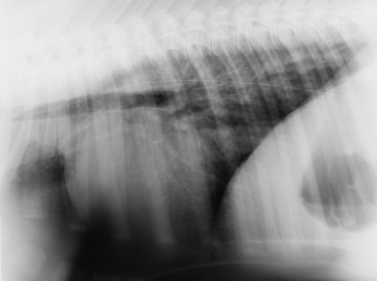
B. Hypoventilation due to prematurity (Figure 45-1)
 |
| Figure 45-1 Right lateral radiograph of a premature foal. A diffuse increased soft tissue opacity partially silhouettes the pulmonary blood vessels (interstitial pattern). This diffuse interstitial lung pattern was caused by prematurity and resolved with no treatment. A tube with a radiopaque marker is present in the esophagus. (From Thrall DE. Textbook of Veterinary Diagnostic Radiology, 5th ed. St Louis, 2007, Saunders.) |
2. Weak muscles
3. Stiff lungs
4. Poor control of pulmonary vasculature
5. Mismatching
6. Shunting
7. Lateral recumbency
8. Atelectasis
9. Pulmonary edema
II. Premature/dysmature foals require
A. Intranasal oxygen
B. Positional support
C. Some require
1. Mechanical ventilation
2. Criteria for using oxygen therapy
a. Pao 2 less than 55 to 60 mm Hg in lateral recumbency
b. Increased RR
c. Labored respirations
d. Increased respiratory and abdominal muscle activity
D. Oxygen toxicity: 100% O 2 for prolonged periods
E. Pulmonary edema, lack of surfactant production, tracheal irritation
INFECTIOUS DISEASES
I. Gram-negative bacteremia with pneumonia (septic foals)
A. Foals younger than 1 week of age
B. Risk factors
1. Prematurity
2. Gestational age greater than 365 days
3. Failure of passive transfer
4. Hypoxemic ischemic encephalopathy (dummy foal)
5. Maternal factors
a. Neonatal isoerythrolysis
b. Unsanitary foaling environment
c. Adverse climatic conditions
C. Gram-negative bacteremia with pneumonia
Stay updated, free articles. Join our Telegram channel

Full access? Get Clinical Tree


