CHAPTER 34. Diagnostic Imaging
Rebecca S. McConnico
CONVENTIONAL RADIOGRAPHY
I. The foot: P3 (distal or 3rd pedal bone)
A. Two views
1. Frontal view: Dorsopalmar, dorsoplantar (DP)
2. Lateral to medial view
B. Anatomy
1. P3 has three surfaces
a. Articular
b. Parietal (face)
c. Solar (bearing surface)
2. Parietal sulcus–dorsopalmar view: Bilateral notches in the lateral cartilages (harbors the dorsal artery of the foot)
3. Solar canal: Pair of small radiolucent ovals in the middle third of the distal phalanx (DP)
4. Solar margin: Appearance is highly variable and should not be mistaken for disease
5. Vascular channels: Size, shape, and number are highly variable
6. Density of P3: Variable and can change with age and use levels
7. Width of the distal interphalangeal joint is often uneven during radiography imaging because of the horse shifting weight
8. Many horses have a notched toe (do not confuse with focal osteomyelitis)
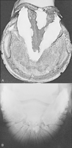
9. If the solar surface is not cleaned and packed before radiography, a characteristic V shape may appear in the high coronary view (which indicates gas in the sulci) (Figure 34-1)
 |
| Figure 34-1 A, The central and collateral sulci of the foot have been packed with a pliable material of similar radiopacity as the sole (Play-Doh). B, Dorsal 65-degree proximal-palmarodistal view of a normal P3 with the sulci packed. Note the good definition of vascular channels without superimposed air artifacts. (From Thrall DE. Textbook of Veterinary Diagnostic Radiology, 5th ed. Philadelphia, 2007, Saunders.) |
C. Abnormalities
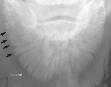
1. Pedal osteitis (Figure 34-2)
 |
| Figure 34-2 Dorsal 65-degree proximal-palmarodistal view of the P3 of a horse with pedal osteitis. Note the irregular margin of the lateral aspect of P3 ( arrows). (From Thrall DE. Textbook of Veterinary Diagnostic Radiology, 5th ed. Philadelphia, 2007, Saunders.) |
a. Nonspecific inflammation of the DP
b. Radiographic signs
(1) Decreased bone density
(2) Course trabeculation
(3) Marginal irregularity
(4) Increased size, shape, and number of vascular channels
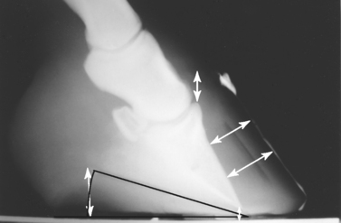
2. Laminitis (Figure 34-3)
 |
| Figure 34-3 Radiograph of a grade III laminitic event. (From Floyd A. Equine Podiatry. Philadelphia, 2007, Saunders.) |
a. Single lateral view is often all that is needed to diagnose distal phalangeal displacement or rotation
b. Not necessary to use metallic devices for reference points because the coronary band is visible
c. Microfractures may be visible at the P3 solar tip
d. Rotation: The distal two thirds of P3 pulls away from the solar laminae, leaving the extensor process in a normal position. Loss of parallel alignment between dorsal hoof wall and dorsal P3
e. Sinking: Linford method, the width of the soft tissue overlying the dorsal surface of P3
f. Gas along the inner surface of the hoof wall constitutes evidence of founder
g. Chronic laminitis: Vascular injury leads to abnormal hoof growth
h. Prognosis
(1) Rotation, distal displacement, and laminar gas result in a poor prognosis
(2) Solar penetration – grave prognosis for soundness
3. Distal phalangeal fractures
a. Common causes
(1) Racing on extensive hard surfaces
(2) Blunt trauma
(3) Preexisting foot disease
(4) Collision with another horse during a race
b. Most commonly affect the articular surface and usually through the left or right lateral surface; may be due to uneven weight distribution
c. Clinical signs
(1) Lameness
(2) Regional hyperemia
(3) Excessive heat
(4) Increased pulses
d. Standard P3 fracture series
(1) High coronary (70-degree displacement)
(2) Right and left frontal obliques
(3) True lateral
e. Distal phalangeal fracture types
(1) Complete extending through the bone in the sagittal or parasagittal plane
(a) Enter the coffin joint
(b) More painful
(2) Solar margin fracture
(a) Toe fractures
(b) Indirect result of distal phalangeal rotation subsequent to laminitis
(c) Marginal sequestrum: Fractures of sequestrum
(3) Wing fractures
(a) Requires oblique views for visualization
(b) Not as painful as articular fractures
(4) Extensor process fractures
(a) Traumatic avulsion
Stay updated, free articles. Join our Telegram channel

Full access? Get Clinical Tree


