The Head
 . Approach to the Rostral Shaft of the Mandible
 . Approach to the Caudal Shaft and Ramus of the Mandible
 . Approach to the Ramus of the Mandible
 . Approach to the Temporomandibular Joint
 . Approach to the Dorsolateral Surface of the Skull
 . Approach to the Caudal Surface of the Skull
Approach to the Rostral Shaft of the Mandible
Based on a Procedure of Rudy40
Description of the Procedure
Additional Exposure
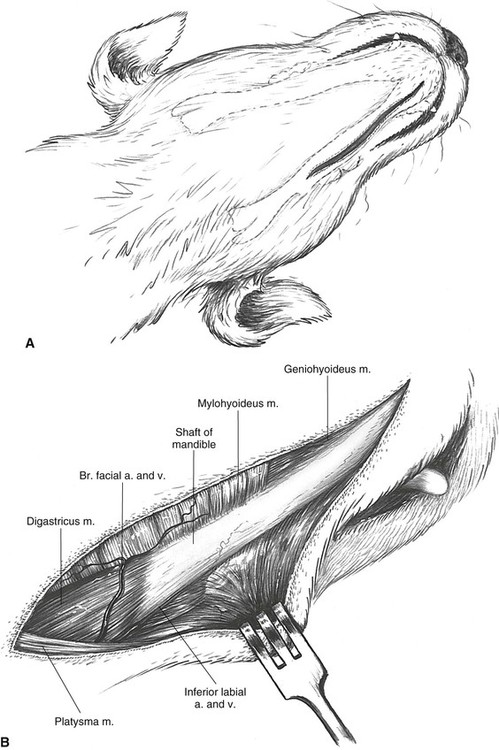
Exposure of the entire shaft of the mandible is obtained by extending the approach more caudally (Plate 7).
Approach to the Caudal Shaft and Ramus of the Mandible
Description of the Procedure
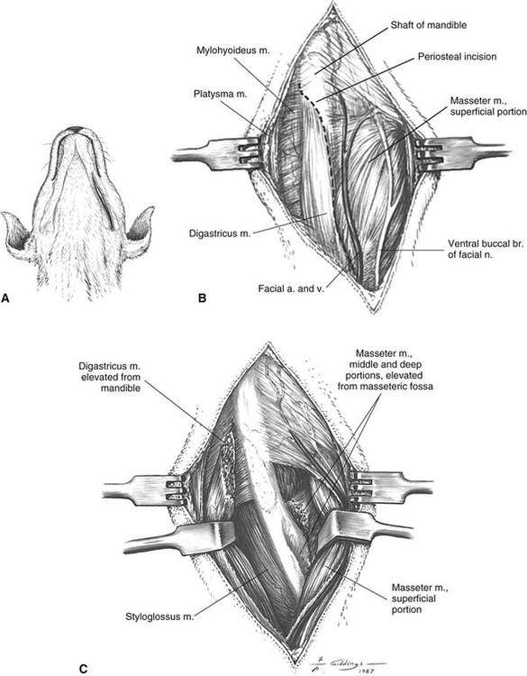
A The incision is centered on the ventral surface of the mandible, commencing at the angular process of the mandible and extending cranially approximately one half the length of the mandible.
B The platysma muscle is incised with the skin to reveal the superficial portion of the masseter muscle laterally and the digastricus muscle lying ventromedially over the shaft of the mandible. An incision is made in the intermuscular septum between the masseter and the digastricus muscles. Lateral to this incision is the large facial vein and accompanying nerve trunks. The periosteal insertion of the digastricus muscle on the shaft of the mandible is incised and elevated.
C Lateral retraction of the masseter and subperiosteal elevation of part of its insertion in the masseteric fossa allow good exposure of the lateral side of the shaft and ventral part of the ramus, and medial retraction of the digastricus and deeper-lying mylohyoideus muscle gives exposure of the medial side of the shaft. The mylohyoideus and rostral insertion of the masseter muscle both can be elevated for more exposure of the ventral border of the mandible.
Additional Exposure
Ventral exposure of the entire shaft and ramus of the mandible is obtained by combining with the approaches described in Plates 6 and 8.
Closure
The intermuscular septum between the digastricus and masseter muscles is closed, with care being taken not to impinge the facial vessels. Platysma muscle is included with subcutaneous fascia in a separate layer.
Stay updated, free articles. Join our Telegram channel

Full access? Get Clinical Tree


