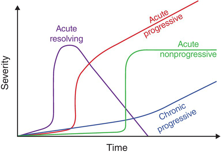
19 James M. Fingeroth and William B. Thomas Clinicians are frequently confronted with patients in which intervertebral disc herniation (IVDH) is considered the working diagnosis. Elsewhere in this text, we have addressed the need for caution in assuming that all dogs that presented with neck pain, back pain, or paresis and ataxia do so because of disc-related disease. We have also touched on (see Chapters 13, 15, and 28) the reasons why there might be a poor correlation between severity of signs and the role that surgery could play in treating those signs. We will expand on that discussion here. The challenge for clinicians is to combine all the information available and recommend treatment that is neither too conservative nor overly aggressive. There are two key concepts that should always be borne in mind when making clinical decisions for dogs and patients with suspected IVDH. First, surgery is only sensible when there is spinal cord compression. As has been demonstrated in other chapters there is a very questionable and probably insignificant value to operations that provide access to the vertebral canal and dural tube/spinal cord, but do not remove a compressive mass. Second, one can neither assume that mild signs (such as discomfort without other deficits) imply minimal spinal cord/nerve root compression, nor the converse that severe signs (tetra or paraplegia) indicate that there must be a significant compressive lesion present. Hence, the clinician uses both inductive and deductive reasoning to decide if a patient might have a compressive lesion for which surgery could be helpful and must further decide when surgery should be considered rather than nonsurgical management. A helpful mental tool in this regard is the construction of a time–sign graph (Figure 19.1). The signs plotted might represent the severity of either discomfort or paresis or both. First, one must consider the historical onset of signs: Did they develop gradually, over days, or longer or were they acute/peracute in onset? Since their initial recognition, how have these signs progressed? Have they stayed the same? Improved? Worsened? If medical intervention was initiated, how has that intervention impacted on the plotted trend? As one considers the onset and progression of signs over time, one can then mentally extrapolate the data to suggest what will transpire over the ensuing time periods. It is from this extrapolation that one can make decisions regarding continuation of medical-only treatment versus referral for possible surgery. In any instance, a trend that suggests either a failure to improve with current treatment and more time or worsening of signs in spite of current treatment would warrant a shift in therapeutic intervention. Moreover, and especially in regard to patients with severe paresis, the goal should always be to intervene aggressively to prevent progression to a state where deep pain perception distal to the lesion is diminished or lost, since (as discussed in Chapter 11) the loss of deep pain is associated with a poorer chance for recovery. Figure 19.1 Time–sign graph. Clinical decision making can be aided by the mental construct of a graph that establishes the onset, severity, and rapidity of progression of clinical signs or deficits. Response (or lack thereof) to medical interventions can be incorporated in the graph. In general, patients at risk for permanent paralysis (correlated with severity of deficits and rapidity of progression), progression of deficits despite appropriate nonsurgical interventions, and failure to improve (even if signs are limited to discomfort alone or mild neurologic deficits) are considered candidates for referral for possible surgical intervention. The urgency of such referral is based on extrapolation of the time–sign graph generated for that patient into the subsequent hours or days, assuming previous trends are maintained. It should be noted that severity of signs at any given time is not as useful in making decisions as is consideration of those signs in the context of onset, progression and response to therapy. Two patients might present with identical signs, but if one reached that point of impairment over a period of days, and the other was normal just a few hours before, it suggests that the latter may have the more emergent condition. It also implies that severity of signs alone is not the only determinant of a need for advanced imaging and surgery. If two patients are presented with, say, signs of severe neck pain but the onset in one occurred just today while the other has been suffering and medically treated for 3 weeks, it is entirely appropriate to consider more aggressive treatment for the latter even though there may yet be no signs of paresis or other neurologic deficits. It is here that we again must remind ourselves that it is quite possible for dogs with signs of discomfort alone to have marked compression of their spinal cords and/or nerve roots, and thus, they are more likely to be benefited from decompressive surgery than from continued medical management [1–3]. This is a common question and one without a simple or definitive answer. However, we can look at some of the pros and cons of plain radiographs, and how the results of such radiography might impinge on treatment recommendations. As noted in Chapter 16, radiographs provide useful clues regarding the presence and site of IVDH. However, as also demonstrated in that chapter, there are many possible artifacts and red herrings that may be seen as well, even when radiographs are done with optimal positioning and technique. There may also be signs of disc degeneration that can occur without corresponding disc herniation (and vice versa), rendering information derived from plain radiographs generally insufficient for a surgeon to base his or her operative planning on. Therefore, the first take-home message is that plain radiographs provided by an attending clinician to a neurosurgeon will, with very rare exception, not obviate the need for subsequent imaging studies. So there should be no motivation for a clinician to obtain plain radiographs as part of a basic database that he or she feels might be helpful to or needed by the neurosurgeon. As with almost any use of radiography, the essential question any clinician should ask is, “How will the results of this imaging alter my diagnostic or treatment strategy for this patient?” As already stated, it is rare for even the best of radiographs to be definitive for making the diagnosis of IVDH. So, if your differential diagnosis list is headed by IVDH, and you consider alternatives to be very unlikely, there is almost no value to having plain radiographs in that circumstance. Your treatment decisions are based on the time–sign graph or similar assessment of the urgency of the case and whether nonsurgical management is or is not appropriate at this time. Hence, if the working diagnosis is IVDH but based on the progression and severity of signs (or the client’s stated intention not to pursue surgery no matter what) you are not currently considering referral for surgery, there is no advantage in having radiographs that may or may not indicate disc disease or even disc herniation. Knowing that there is a narrowed disc space at, say, T13–L1 versus L2–L3 will have no impact on treatment planning or efficacy. And considering the added cost for radiography and morbidity involved (either restraining an anxious patient or those in pain or anesthetizing the patient), it might be considered detrimental to pursue plain radiographs. That all said, we do have to remember that a putative diagnosis of IVDH is just that, and so an argument can be made for using radiographs to screen for the presence of nondisc-related lesions. These can include fractures/luxations (although, in most cases, the history will indicate if external trauma should be considered in the differential diagnosis), discospondylitis (Chapter 20), and vertebral or disc neoplasia (Chapter 21). However, the sensitivity for each of these alternate diagnoses via plain radiography is less than 100%, so negative plain radiographs (especially if obtained with less than optimal positioning, technique, or orthogonal views) cannot rule out any diagnosis. Many patients may also have developmental/anomalous lesions such as hemivertebrae, block vertebrae, butterfly vertebrae, extra or missing vertebrae, or degenerative lesions such as spondylosis deformans, diffuse idiopathic skeletal hyperostosis (DISH), or mineralized discs that may have no relation to current signs of discomfort and/or paresis. So the value of plain radiographs to rule in or rule out alternative diagnoses to IVDH, just as with trying to prove IVDH via plain radiography, is questionable. However, if referral for advanced imaging/surgery entails a great distance or other major logistical hurdle for the client, it can be logically argued that plain radiographs for screening, even in the face of possible false negatives and false positives, should be considered, since a definitive finding of pathology other then IVDH might alter the plans for referral. As a general rule, however, the foregoing should at least sway clinicians from a “knee-jerk” policy of diagnosing a patient with neck or back disease, assuming it to be IVDH, and automatically taking radiographs. If a patient is to be classified a surgical candidate, that classification is based on the differential diagnosis (could there be a compressive lesion affecting the spinal cord and/or nerve roots, be it disc or something else?), the consideration of severity of signs, progression over time, anticipated progression/course without surgery, and the desire to avoid loss of deep pain. Client willingness to pursue referral for possible surgery and response to any medical intervention already initiated are also important considerations. Once the clinical decision is made to recommend referral for surgery, it is only then that imaging studies need to be performed. There are virtually no instances where the results of radiography in the primary clinician’s facility or emergency clinic will or should instigate a recommendation for surgery if no such recommendation was being considered before those radiographs were obtained. And if referral for surgery is deemed warranted, any surgical intervention will be predicated on an advanced imaging modality (Chapter 16), making radiographs prior to referral essentially useless, except for the screening motive addressed earlier in instances where the referral might be a logistical hardship for the client.
When Should Dogs Be Referred for Imaging and Surgery?

Should I take plain radiographs of patients in which I suspect have IVDH?
Stay updated, free articles. Join our Telegram channel

Full access? Get Clinical Tree


