Chapter 5. Vitamins
A general classification scheme for vitamins divides them into two groups: the fat-soluble vitamins and the water-soluble vitamins. The fat-soluble vitamins are A, D, E, and K; the water-soluble group includes vitamin C and members of the B-complex vitamin group. Fat-soluble vitamins are digested and absorbed using the same mechanisms as dietary fat, and their metabolites are excreted primarily in the feces through the bile. In contrast, most of the water-soluble vitamins are absorbed passively in the small intestine and are excreted in the urine. Excesses of fat-soluble vitamins are stored primarily in the liver. With the exception of cobalamin, the body is unable to store significant levels of the water-soluble vitamins. As a result, the fat-soluble vitamins, specifically vitamins A and D, have a much higher potential for toxicity than do the water-soluble vitamins. Similarly, because they can be stored, deficiencies of fat-soluble vitamins develop much more slowly in animals than do deficiencies of the water-soluble vitamins. A summary of food sources and signs of deficiency and excess for the vitamins is given in Table 5-1.
| CNS, Central nervous system. | |||
| V itamin | D eficiency | E xcess | S ources |
|---|---|---|---|
| A | Impaired growth, reproductive failure, loss of epithelial integrity, dermatoses | Skeletal abnormalities, hyperesthesia | Fish liver oils, milk, liver, egg yolk |
| D | Rickets, osteomalacia, nutritional secondary hyperparathyroidism | Hypercalcemia, bone resorption, soft tissue calcification | Liver, some fish, egg yolk, sunlight |
| E | Reproductive failure, pansteatitis in cats | Nontoxic; may increase vitamin A and D requirements | Wheat germ, corn and soybean oils |
| K | Increased clotting time, hemorrhage | None recorded | Green leafy plants, liver, some fish meals |
| Thiamin | CNS dysfunction, anorexia, weight loss | Nontoxic | Meat, wheat germ |
| Riboflavin | CNS dysfunction, dermatitis | Nontoxic | Milk, organ meats, vegetables |
| Niacin | Black tongue disease | Nontoxic | Meat, legumes, grains |
| Pyridoxine | Microcytic hypochromic anemia | None recorded | Organ meats, fish, wheat germ |
| Pantothenic acid | Anorexia, weight loss | None recorded | Liver, kidney, dairy products, legumes |
| Biotin | Dermatitis | Nontoxic | Eggs, liver, milk, legumes |
| Folic acid | Anemia, leukopenia | Nontoxic | Liver, kidney, green leafy vegetables |
| Cobalamin | Anemia | Nontoxic | Meat, fish, poultry |
| Choline | Neurological dysfunction, fatty liver | Diarrhea | Egg yolk, organ meats, legumes, dairy products |
| C | Not required by dogs and cats | Nontoxic | Citrus fruit, dark green vegetables |
The fat-soluble vitamins are A, D, E, and K; the water-soluble group includes vitamin C and members of the B-complex vitamin group. Fat-soluble vitamins are digested and absorbed using the same mechanisms as dietary fat, and their metabolites are excreted primarily in the feces through the bile. Most of the water-soluble vitamins are absorbed passively in the small intestine and are excreted in the urine.
VITAMIN A
The general term vitamin A actually includes several related chemical compounds called retinol, retinal, and retinoic acid. Of these molecules, retinol is the most biologically active form. In the body, vitamin A has functions involving vision, bone growth, reproduction, and maintenance of epithelial tissue. The role of vitamin A in vision is well established. In the rods of the retina, retinal combines with a protein called opsin to form rhodopsin, also known as visual purple. Rhodopsin is a light-sensitive pigment that enables the eye to adapt to changes in light intensity. When exposed to light, rhodopsin splits into retinal and opsin, and the energy that is released produces nerve transmissions to the optic nerve. In the dark, rhodopsin can then be regenerated by the combination of new retinal and opsin molecules. During periods of vitamin A deficiency, less retinal is available to regenerate rhodopsin; thus the rods of the eye become increasingly sensitive to light changes, which eventually results in night blindness.
Vitamin A is also essential for the formation and maintenance of healthy epithelial tissue. This tissue includes the skin and the mucous membranes lining the respiratory and gastrointestinal tracts. Vitamin A is believed to be necessary for both the proliferation and differentiation of cells and for the production of the mucoproteins found in the mucus produced by some types of epithelial cells. 1 Mucous secretions of epithelial tissue maintain the integrity of the epithelium and provide a barrier against bacterial invasion. In the absence of vitamin A, the differentiation of new epithelial cells beyond the squamous type to mature mucus-secreting cells fails to occur, and normal epithelial cells are replaced by dysfunctional, stratified, keratinized cells. 2 Epithelial tissue that does not function properly leads to lesions in the epithelium and increased susceptibility to infection.
Normal skeletal and tooth development and reproductive performance also depend on vitamin A. The vitamin’s role in bone growth appears to involve the activity of the osteoclasts and osteoblasts of the epithelial cartilage and may be related to cellular division and the maintenance of cell membranes through glycoprotein synthesis. Experiments with laboratory animals have shown that vitamin A is also essential for spermatogenesis in males and normal estrous cycles in females. 1
The origin of all vitamin A is the carotenoids, which are synthesized by plant cells. Carotenoids are dark red pigments that provide the deep yellow/orange color of many plants. Vegetables such as carrots and sweet potatoes contain high amounts of these compounds. Deep green vegetables also contain these pigments, but their color is masked by the deep green color of chlorophyll. When animals consume the carotenoids in plants, an enzyme located in the intestinal mucosa, β-carotene 15,15′-dioxygenase, converts these compounds (which are commonly called provitamin A) to active vitamin A (Figure 5-1). The active vitamin is then absorbed and stored principally in the liver. Although several different carotenoids are capable of providing vitamin A, beta-carotene is the most plentiful in foods and has the highest biological activity. Animal products do not contain carotenoids but can provide active vitamin A when included in the diet. Fish liver oils contain the highest amounts, and more common foods such as milk, liver, and egg yolk also contain vitamin A.
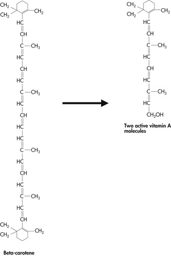 |
| Figure 5-1 |
Like most animals, dogs are capable of converting carotenoids to active vitamin A; therefore they do not require an animal source of this vitamin in the diet. However, β-carotene 15,15′-dioxygenase is either absent or deficient in the domestic cat. As a result, the cat is unable to convert carotenoid pigments to vitamin A and must receive a source of preformed vitamin A in the diet (see Section 2, pp. 107-108 for a complete discussion). In addition to providing a source of vitamin A, carotenoid pigments also have a role in modulating immune response. Recent studies have shown that both dogs and cats readily absorb beta-carotene and a related carotenoid called lutein, and that these pigments may have a function in cell-mediated and humoral immune response in these species. 3.4. and 5. Vitamin A may also have a role in weight maintenance.
VITAMIN D
Vitamin D consists of a group of sterol compounds that regulate calcium and phosphorus metabolism in the body. As with vitamin A, there are provitamin forms of this vitamin. These are vitamin D 2 (ergocalciferol) and vitamin D 3 (cholecalciferol). Vitamin D 2 is formed when the compound ergosterol, which is found in many plants, is exposed to ultraviolet (UV) radiation. This conversion occurs only in harvested or injured plants, not in living plant tissue. Therefore this form of vitamin D is only of significance to ruminants and nonruminant herbivores that are consuming sun-dried or irradiated plant materials. In addition, most species, including cats, use ergocalciferol less efficiently than cholecalciferol. 6 The second form of provitamin D, vitamin D 3, is the form that is of greatest nutritional importance to omnivores and carnivores such as the dog and cat. It is synthesized by the body when 7-dehydrocholesterol, a compound found in the skin of animals, is exposed to UV light from the sun. This form of vitamin D can be obtained either through synthesis in the skin or from the consumption of animal products that contain cholecalciferol. Dogs and cats appear to be dependent upon dietary sources of vitamin D because they have limited ability to convert 7-dehydrocholesterol in the skin to cholecalciferol. 7. and 8.
Because active vitamin D is synthesized by the body and because of the regulatory functions that it performs within the body, some controversy exists regarding its classification. Although some scientists believe that vitamin D should be considered a hormone, others continue to classify it as a vitamin. Regardless of its categorization, precursors of vitamin D are obtained through the diet, and vitamin D’s functions are intricately involved with normal calcium and phosphorus homeostasis in the body. Both ingested and endogenous vitamin D 3 (cholecalciferol) are stored in liver, muscle, and adipose tissue. Cholecalciferol is an inactive storage form of vitamin D. To become active, it must first be transported from the skin or intestines to the liver; there it is hydroxylated to 25-hydroxycholecalciferol. This compound is then transported through the bloodstream to the kidneys, where it is further converted to one of several possible metabolites. Metabolites include 1,25-dihydroxycholecalciferol, also called calcitriol, which is the most active form of vitamin D (Figure 5-2). The conversion of 25-hydroxycholecalciferol to calcitriol in the kidneys occurs in response to elevated parathyroid hormone (PTH), which is released from the parathyroid gland in response to decreasing serum calcium. A decrease in serum phosphorus also stimulates the formation of active vitamin D in the kidneys. Although inactive vitamin D is considered a vitamin, calcitriol is often classified as a hormone because it is produced in the body and because of its mechanism of action in the body.
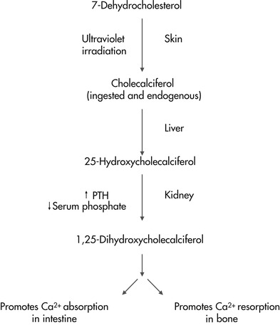
Active vitamin D functions in normal bone tissue development and maintenance and is an important component in the homeostasis of the body’s calcium and phosphorus pools. These effects are mediated through the influence of vitamin D on calcium and phosphorus absorption from the gastrointestinal tract and their deposition in bone tissue. At the site of the intestine, vitamin D stimulates the synthesis of calcium-binding protein, which is necessary for the efficient absorption of dietary calcium and phosphorus. Vitamin D also affects normal bone growth and calcification by acting with PTH to mobilize calcium from bone and by causing an increase in phosphate reabsorption in the kidneys. The net effect of vitamin D’s actions in intestines, bones, and kidneys is an increase in plasma calcium and phosphorus to the level that is necessary to allow for the normal mineralization and remodeling of bone. A deficiency of vitamin D causes impaired bone mineralization and results in osteomalacia in adult animals and rickets in growing animals (see Section 2, pp. 108-110).
Dietary sources of vitamin D for dogs and cats are varied. In general, most natural food substances contain very little vitamin D, although egg yolks, liver, and certain types of fish contain moderate amounts. Among the few concentrated food sources of vitamin D are the fish liver oils, particularly cod liver oil. Because natural foods are low in this vitamin, most commercially prepared pet foods are enriched with a purified form of cholecalciferol (vitamin D 3).
VITAMIN E
Vitamin E is the term used to describe a group of chemically related compounds called the tocopherols and tocotrienols, which have varying levels of biological activity relative to the most potent form, alpha-tocopherol. 9 There are four naturally occurring tocopherols; of these, alpha-tocopherol is the most active form of vitamin E and is the compound most commonly included in pet foods. Several active synthetic forms of vitamin E have also been produced and are used in processed foods. Within the body, vitamin E is found in at least small amounts in almost all tissues and is incorporated into the membrane bilayer of cells. The liver is able to store appreciable amounts of vitamin E.
< div class='tao-gold-member'>
Only gold members can continue reading. Log In or Register to continue
Stay updated, free articles. Join our Telegram channel

Full access? Get Clinical Tree


