Chapter 9 THE VERTEBRAL COLUMN
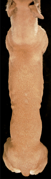
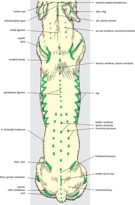
Fig. 9.1 Surface features of the neck, trunk and tail: dorsal view. The palpable bony ‘landmarks’ of the vertebral column and adjoining bones are shown in this figure. For comparable lateral views see Figs 3.1, 5.1, 6.1 and 7.6.
Fig. 9.2 Axial skeleton: dorsal view. The palpable bony features shown in the surface view are colored green for reference. Other bony features of the vertebral column are normally impalpable through the overlying dorsal (epaxial) musculature, especially in the neck and lumbar region. For comparable lateral views of the skeleton see Figs 3.2, 5.2, 6.2 and 7.7.
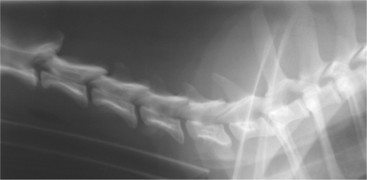
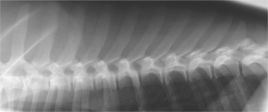
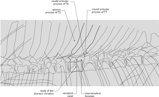
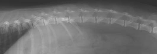
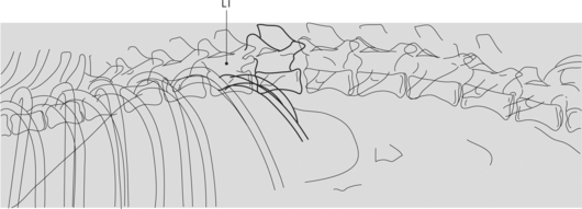
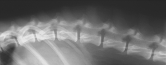
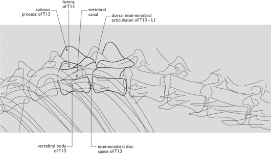
Fig. 9.7 Radiograph of the thoracolumbar junction: lateral view. The intervertebral synovial articulations are orientated in the sagittal plane from the anticlinal vertebra caudally (compare with Figure 9.5).
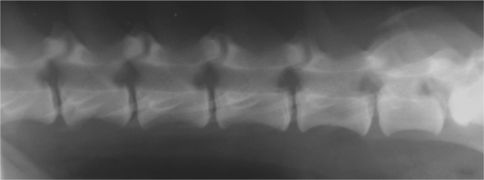
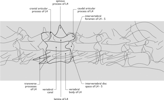
Fig. 9.8 Radiograph of the lumbar spine: lateral view. The large intervertebral foramina can be clearly seen.
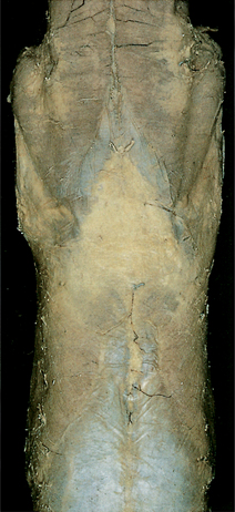
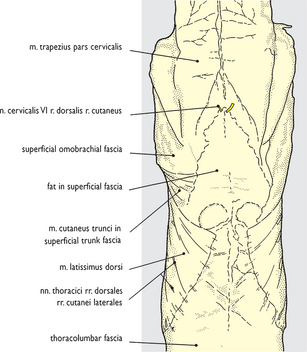
Fig. 9.9 Superficial structures of the caudal end of the neck and thoracic vertebral region: dorsal view. The loose superficial fascia is variably infiltrated with fat and contains the cutaneous muscle of the trunk. Lateral cutaneous branches from the dorsal rami of thoracic nerves are apparent in the dorsolateral thorax. Comparable views from the lateral aspect are shown in Figs 3.6 and 5.7.
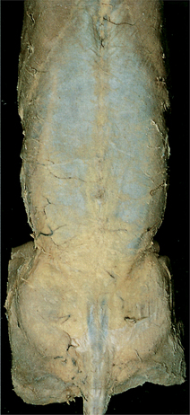
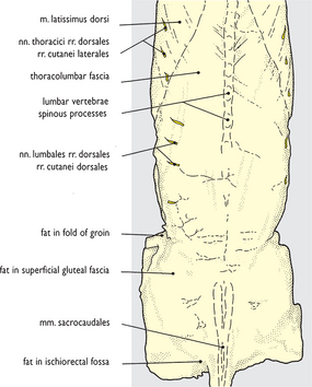
Fig. 9.10 Superficial structures of the lumbar and pelvic regions and the root of the tail: dorsal view. Fat is quite extensively infiltrated into the lumbar and gluteal fascia and is present in the fold of the flank and ischiorectal fossa. Comparable views from a lateral aspect are shown in Figs 6.11 and 7.11.
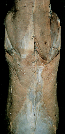
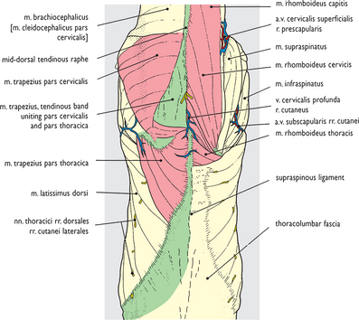
Fig. 9.11 Superficial muscles of the caudal end of the neck and the thoracic vertebral region: dorsal view. The superficial fascia has been cleaned from the surface to expose superficial extrinsic limb muscles on the left side. These are not covered externally by the deep thoracolumbar fascia which is continued internal to them on the surface of the epaxial musculature. On the right side the cleidocervical component of the brachiocephalic and the trapezius muscles have been removed to expose the rhomboid muscle in the interscapular region. For comparable views from the lateral aspect see Figs 5.9 and 5.10.
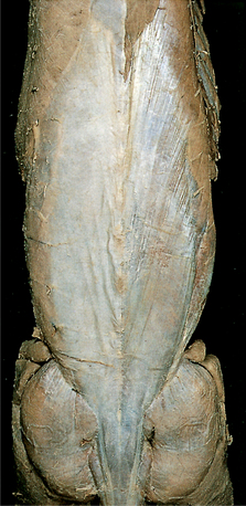
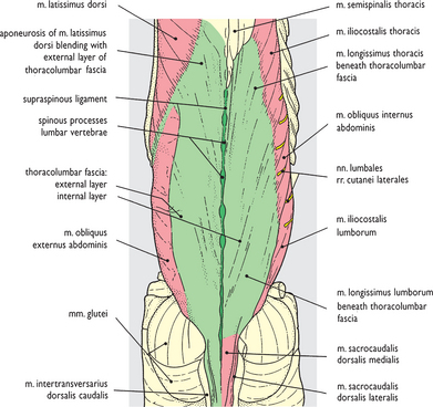
Fig. 9.12 Superficial muscles of the lumbar and pelvic regions and the root of the tail: dorsal view. Cleaning of superficial fascia from the surface has exposed the superficial muscles to some extent, although the dense thoracolumbar fascia obscures detail of the underlying epaxial muscles. On the right side the external layer of thoracolumbar fascia has been removed along with the latissimus dorsi and external abdominal oblique muscles, exposing an internal layer of fascia closely applied to the muscles. For comparable views from the lateral aspect see Figs 6.14, 6.15 and 7.15.
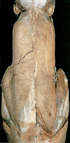
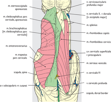
Fig. 9.13 Superficial muscles of the neck and interscapular region: dorsal view. The superficial fascia has been cleaned from the left side of the neck and shoulder to expose the superficial muscles. This removal also included the platysma muscle (see Figs 3.6 and 3.7) and the muscles of the auricular cartilages (see Figs 2.41–2.45). On the right side removal of the cleidocervical component of the brachiocephalic muscle and the cervical trapezius has exposed the extent of the cervical and capital parts of the rhomboid muscle. Comparable views of the neck from a lateral aspect are given in Figs 3.13 and 3.15.
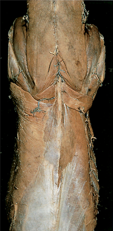
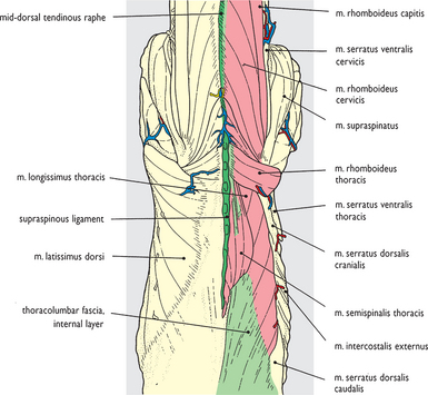
Fig. 9.14 Rhomboid and thoracic epaxial muscles after removal of the trapezius and latissimus dorsi muscles: dorsal view. Removal of the latissimus dorsi muscle on the right side completes the exposure of the thoracic component of the rhomboid muscle in the interscapular region. The thoracolumbar fascia is continued cranially on the surface of the epaxial muscles beneath the thoracic rhomboid; through it the combined spinal and semispinal muscle of the thorax and the thoracic longissimus are visible. A comparable view from the lateral aspect is given in Fig. 5.10 in which the deep thoracolumbar fascia between the attachments of the dorsal serrate muscles has been removed.


