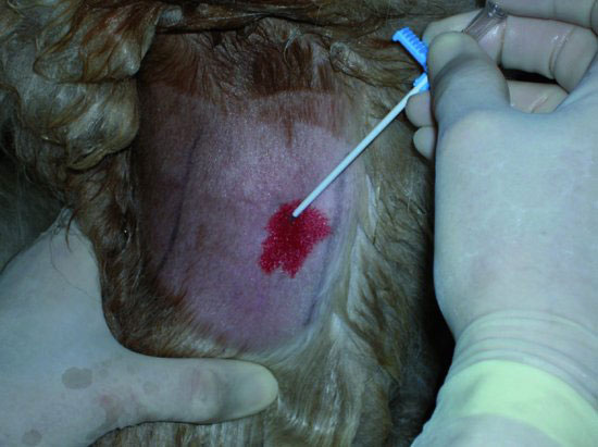
Figure 12.2 Injection of 2% lidocaine for local anesthesia subcutaneously over the jugular vein prior to catheterization.
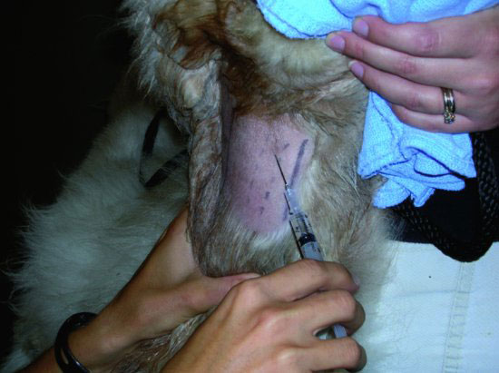
Figure 12.3 Stab incision made with a #15 or #11 scalpel blade through the skin overlying the jugular vein. This can be helpful in minimizing catheter drag.
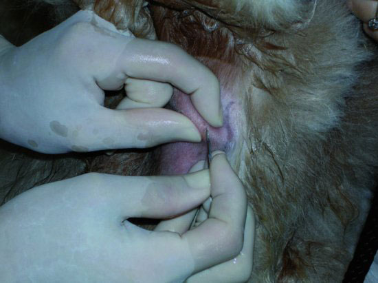
Figure 12.4 Occlusion of the jugular vein with the veterinarian’s left hand with placement of the catheter/stylette unit into the jugular vein.
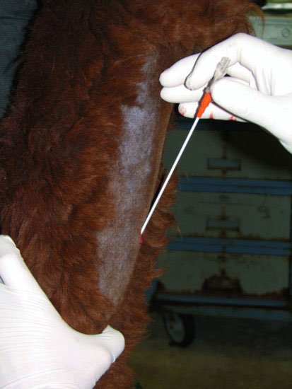
Figure 12.5 The catheter has been introduced fully into the jugular vein and the stylette removed. The vein is occluded to confirm placement.
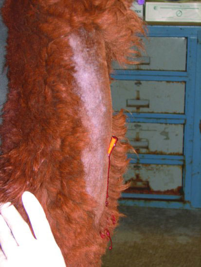
Figure 12.6 Attachment of an extension set to the catheter for ease of bandaging and fluid administration.
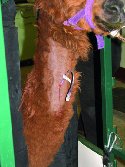
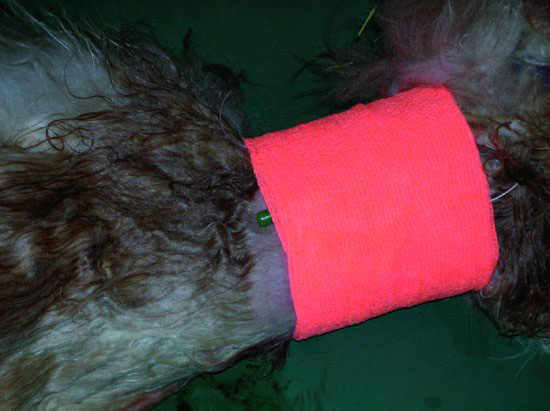
Figure 12.7 A neck wrap placed over a jugular catheter. The injection port is left outside the wrap for easy access.
Camelid “peel away” catheters (Figures 12.8–12.13) and J-wire catheters (Figures 12.14–12.22) require additional steps for placement as compared to over-the-needle catheters, and step-by-step photos are provided for these.
Figure 12.8 The package contents of the camelid “peel away” catheters, which includes a syringe, catheter, and insertion catheter and stylette.
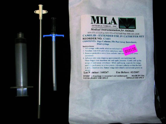
Figure 12.9 Insertion of the stylette and tabbed insertion catheter into the jugular vein.
Stay updated, free articles. Join our Telegram channel

Full access? Get Clinical Tree


