48 VAGINAL CYTOLOGY
2 What are basal cells?
Basal cells are located on the basement membrane and are the precursor cell for the other vaginal epithelial cells (parabasal, intermediate, superficial). Basal cells are seldom observed on vaginal cytology smears and appear as small cells with a small amount of basophilic cytoplasm.
3 What are parabasal cells?
Parabasal cells are the smallest and most immature of the epithelial cells typically observed on cytology smears. They appear as small round cells with a small amount of basophilic cytoplasm and a roundish nucleus. Parabasal cells with cytoplasmic vacuoles are called foam cells.
4 What are intermediate cells?
Intermediate cells are larger than parabasal cells, often being twice as large or larger. Depending on their size, intermediate cells are subclassified into small and large intermediate cells. The size of intermediate cells depends on the amount of their cytoplasm because the nucleus of both large and small intermediate cells is about the same size and typically roundish. The cytoplasm of small intermediate cells is usually smooth and round to slightly oval. The cytoplasm of large intermediate cells tends to be angular and occasionally may also be irregular and folded. The cytoplasm is typically blue to blue-green in both small and large intermediate epithelial cells. The terms superficial and transitional are occasionally used for large intermediate cells.
5 What are superficial cells?
Superficial cells are the most mature cells of vaginal epithelial stages. They are large cells with abundant, angular or folded, blue to blue-green cytoplasm. The cytoplasm may contain a few to many vacuoles, and some cells may contain dark-staining bodies of unknown significance. The nucleus is smaller than that of intermediate cells and may be faded or even absent (anucleate). The maturation process and development of superficial cells are often referred to as cornification, and superficial epithelial cells are sometimes referred to as cornified cells.
7 What are the cytologic features of proestrus in dogs?
8 Are the red blood cells observed on proestrus vaginal cytology smears from vaginal vessels?
RBCs observed during proestrus are not from vaginal vessels; they are erythrocytes exiting uterine capillaries by diapedesis.
9 What is the duration of proestrus in dogs?
In normal dogs, proestrus ranges from 2 to 17 days (depending on the reference source) with a mean duration of 9 days.
10 How do the cytologic features of proestrus in cats differ from those in dogs?
In cats, RBCs and neutrophils are absent, so only the epithelial cells are present. The epithelial cell changes are similar to those in dogs. Also, bacteria may be present.
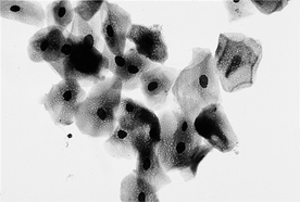
11 What are the cytologic features of estrus in dogs?

Figure 48-1 Estrus. Vaginal smear from a dog consisting of approximately 100% large superficial epithelial cells. (Wright’s stain; 125×.)

Stay updated, free articles. Join our Telegram channel

Full access? Get Clinical Tree

