Uveitis, Equine Recurrent

Basic Information 
Clinical Presentation
Disease Forms/Subtypes
Three main clinical syndromes are observed in ERU: classic, insidious, and posterior.
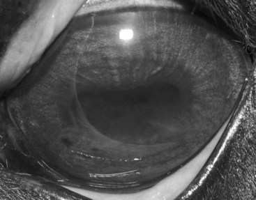
• Classic type: Most common and is characterized by active inflammatory episodes in the eye followed by periods of minimal ocular inflammation. The acute, active phase of ERU predominantly involves inflammation of the iris, ciliary body, and choroid, with concurrent involvement of the cornea, anterior chamber, lens, retina, and vitreous. After treatment with nonspecific antiinflammatory medications such as corticosteroids, the signs of active, acute uveitis can recede, and the disease enters a quiescent or chronic phase. After variable periods of time, the quiescent phase is generally followed by further and increasingly severe episodes of uveitis. The recurrent, progressive nature of the disease is responsible for development of cataracts, intraocular adhesions, and phthisis bulbi (scarred eye) (Figure 1).
• Insidious type: The inflammation never completely resolves, and a low-grade inflammatory response continues that leads to chronic clinical signs of ERU. Frequently, these horses do not demonstrate overt ocular discomfort, and owners of these horses may not recognize the presence of disease until a cataract forms or the eye becomes blind. This type of uveitis is most commonly seen in Appaloosa and Draft Horses.
• Posterior type: Has clinical signs existing entirely in the vitreous and retina, with little or no anterior signs of uveitis. In this syndrome, there are vitreal opacities and retinal inflammation and degeneration.
History, Chief Complaint
Persistent or recurrent blepharospasm, epiphora, ocular cloudiness, and blindness
Physical Exam Findings
• Typical clinical signs of active RU are similar to signs of uveitis in other species: Photophobia, blepharospasm, corneal edema, aqueous flare, hypopyon, miosis, vitreous haze, and chorioretinitis.
• Clinical signs of chronic RU include corneal edema, iris fibrosis and hyperpigmentation, posterior synechia, corpora nigra degeneration (smooth edges), miosis, cataract formation, vitreous degeneration and discoloration, and peripapillary retinal degeneration (Figure 2).
• The classic and insidious types of RU can have either predominantly anterior (cornea, iris, lens, and ciliary body inflammation) or posterior (ciliary body, vitreous, and chorioretinal inflammation) segment involvement. Ultimately, even with aggressive treatment, many horses develop a chronically painful eye and blindness as a result of secondary cataract, synechia (intraocular adhesions), scarring, glaucoma, and development of phthisis bulbi.
Stay updated, free articles. Join our Telegram channel

Full access? Get Clinical Tree


