39 UMN
Spinal cord infarction
INTRODUCTION
Spinal cord disease may present with either upper motor neuron (UMN) or lower motor neuron (LMN) signs, or a combination of the two, depending on the location of the lesion.
CASE HISTORY
This dog began dragging the dorsum of the right hind paw. Later the same day the right forelimb started to collapse when weight-bearing. The dog seemed a little depressed but brightened up after pain relief was given by the local vet. There had been no prior illness or injury reported. The signs had not apparently improved or worsened during the 3 days prior to referral.
CLINICAL EXAMINATION
The dog was alert and non-ambulatory. When held up, voluntary movement was seen in all limbs. Hopping was normal in the left forelimb, slow in the left hind, and absent in the right fore and hind limbs. Spinal reflexes were normal to increased in all limbs. Muscle tone was increased in all limbs. No spinal pain was elicited.
NEUROANATOMICAL LOCALIZATION
The lesion was localized to the cervical spinal cord segments C1–C5, on the right hand side.
DIFFERENTIAL DIAGNOSIS
The sudden onset and strikingly lateralized nature of the signs of this spinal cord disease are very suggestive of spinal cord infarction caused by fibrocartilagenous thromboembolism. Compressive cord disease such as intervertebral disc disease (IVDD) and neoplasia is less focal as a general rule and tends to progress. Sudden onset IVDD is usually painful.
CASE WORK-UP
MRI of the cervical cord 4 days after the onset of signs found an area of hyperintensity on T2WI within the right side of the spinal cord at the level of the C4–5 vertebrae. CSF was normal, but this may have reflected sampling rostral to the lesion (Fig. 39.1).
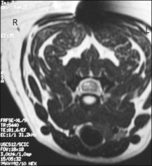
Figure 39.1 Transverse T2WI. Focal hyperintensity within the right side of the cervical spinal cord.
Haematology and biochemistry were normal. Clinical signs had improved overnight and the animal was ambulatory with proprioceptive deficits of the right fore and hind paws at each step.
DIAGNOSIS
The diagnosis is based on a combination of factors as no one factor is diagnostic in itself. The sudden onset, progression, lateralization and improvement, coupled with signal changes on MRI suggestive of intramedullary oedema or tissue necrosis supported the clinical diagnosis of a fibrocartilagenous thromboembolism.
Stay updated, free articles. Join our Telegram channel
Full access? Get Clinical Tree
