Figure 50.2 Image of the right kidney showing the smooth capsular appearance (C), the renal cortical parenchyma (A), and pelvis (B).
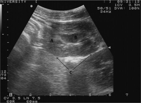
Figure 50.3 Approximate probe positioning for imaging the left kidney is more caudal than the right. By positioning the probe face cranial to the left kidney, the spleen can be located.
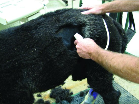
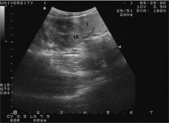
Figure 50.4 An ultrasound image of the left kidney (LK), showing the close approximation to the spleen (S). Splenic vasculature can be seen coursing through the parenchyma.
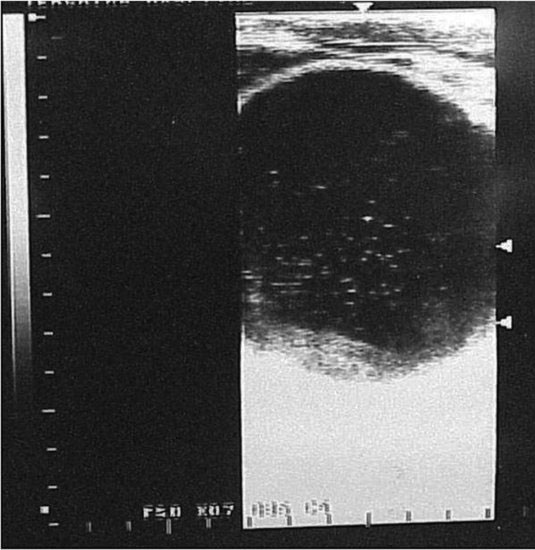
Place the probe in the caudoventral flank and direct it caudodorsally to the pelvis, to allow visualization of the urinary bladder. If the bladder is empty, it will likely not be visualized. Severe distention is easily recognized in cases of urethral obstruction as might be seen with urolithiasis. (See Figure 50.5.) The normal bladder should not be more than 6 to 8 cm in diameter in the adult camelid or appear very tense. Hyperechoic irregularities may be seen suspended in the bladder lumen of normal patients and are not pathognomonic of uroliths or mucosal debris. However, with overdistention and a compatible history of urethral obstruction or cystitis, these refractile bodies may be pathologic.
Figure 50.5 Transabdominal ultrasound image showing an overdistended urinary bladder. The measurement scale to the left is in centimeters. Note the refractile bodies within the lumen.
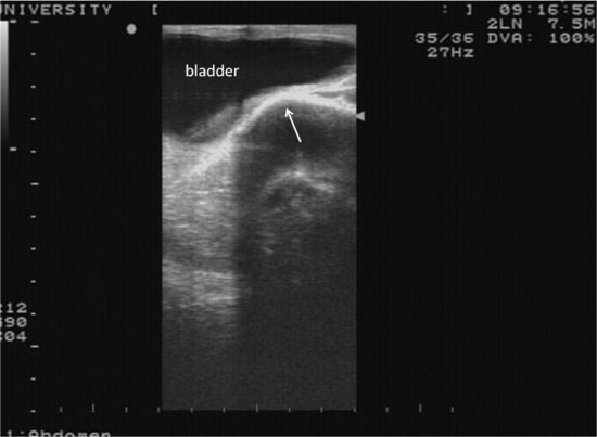
Rectal ultrasound provides detailed images of the bladder wall, trigone, pelvic urethra, and accessory sex glands in males (Figure 50.6). A form of prostatic hyperplasia and prostatic cysts causing stranguria has been noted by this author in young males, and the occurrence has been briefly mentioned by others (Figure 50.7) (Tibary and Vaughan, 2006). Another problem seen in camelids is a form of idiopathic polypoid cystitis (Figure 50.8). To date, biopsy of these lesions has only reported chronic inflammation without specific causes. Some cases resolve with medical management, where others require surgery (Anderson et al., 2010). Bladder wall masses may involve both the serosal and mucosal surfaces and vary in size and number (Figure 50.9). Although neoplasia from suspected bracken fern toxicosis has been reported in a llama, lesions were isolated to the kidneys and ureters. Gross bladder lesions were not evident as would be expected in cattle (Peauroi, et al. 1995).
Figure 50.6 An ultrasound image of a normal urinary bladder as seen per the rectum at the level of the pubic eminence (arrow).
Stay updated, free articles. Join our Telegram channel

Full access? Get Clinical Tree


