15 Tumors of the Nasal and Paranasal Sinuses
Most tumors in the nasal cavity of the dog are malignant. They typically originate from the epithelium lining the nasal and paranasal cavities. Adenocarcinomas are most frequent; while mesenchymal cell tumors (fibrosarcomas, chondrosarcomas, and osteosarcomas) are less frequently diagnosed. Unusual tumors are mast cell and transmissible venereal tumors.
On average, dogs with nasal cancer are about 10 years old, but nasal tumors have been reported in dogs younger than 1 year. Chondrosarcomas occur in younger dogs (mean, about 7 years old). Males may be more frequently affected. Dolichocephalic and mesocephalic breeds are at increased risk compared to brachycephalic (short-nosed) breeds, possibly because in dolichocephalic breeds more of the nasal turbinate surface is exposed to carcinogens. Living in urban areas and exposure to flea sprays, by-products of indoor kerosene/coal-burning heating units, and second hand tobacco smoke are associated with an increased risk of developing nasal neoplasms.
The most common presenting clinical signs are epistaxis, unilateral or bilateral nasal discharge, airflow obstruction, stridor, facial deformity, and sneezing. Less frequent findings include dyspnea, coughing, weight loss, tonsillitis, exophthalmos, and reverse sneezing. These signs have often been present for 3 to 4 months prior to presentation to a veterinarian. The response to antimicrobial and/or corticosteroid therapy is often transient. Invasion of the cribriform plate causes neurologic abnormalities (seizures, paresis) and may be seen in dogs without any clinical evidence of nasal or paranasal sinus involvement. Paraneoplastic disorders are rare.
The differential diagnoses for chronic nasal discharge include bacterial rhinitis; fungal rhinitis (aspergillosis, penicilliosis, rhinosporidiosis, cryptococcosis, and pythiosis); inflammatory disorders (lymphoplasmacytic or eosinophilic rhinitis); parasitic rhinitis (Pneumonyssoides caninum), foreign body (plant awn); abscess of a tooth root with drainage into the nasal cavity; and inflammatory polyp. Depending on the nature of the vessel destruction, epistaxis can occur alone or with a serous or mucopurulent nasal discharge. Nonrespiratory tract causes of epistaxis include disorders of primary hemostasis (von Willebrand’s disease), immune-mediated thrombocytopenia, canine ehrlichiosis, coagulopathies, and vascular disorders (hypertension, hyperviscosity syndrome).
The complete workup for a dog with a nasal discharge typically involves radiographs, computed tomography (CT), or magnetic resonance imaging (MRI) and biopsy (either blind or with rhinoscopy). Plain radiography will help confirm the presence of nasal/paranasal sinus disease but often does not help differentiate among infectious, inflammatory, and neoplastic diseases. Evaluation of nasal exudates obtained by swabbing or a saline solution flush is most often unrewarding. Aerobic culturing generally results in an abundant yield of normal bacterial flora or bacterial pathogens representing a secondary infection that accompanies the primary disease process. Fungal infections may be differentiated from neoplasia, bacterial infection, and allergic nasal disease with a combination of radiographic signs, rhinoscopic examination, and serologic examination. Cryptococcus organisms may be easily identified on cytologic examination of exudates; however, a cytologic diagnosis of nasal cancer from exudate examination alone is made infrequently. Fungal hyphae (Aspergillus spp.) may be observed in the exudates of healthy dogs. Penicillium and Aspergillus may be grown in cultures from healthy dogs or dogs with a variety of other nasal diseases. Unfortunately, fungal serology is only reliably useful for Cryptococcus.
Histopathologic examination of tissue from the nasal cavity is often the only way to differentiate between the varieties of nasal diseases, particularly nasal cancer. Few laboratory abnormalities are associated with the most common nasal tumors of dogs. On careful oral examination, palpation may reveal a mass in the pharynx or deviation of the hard palate or signs of dental disease may be noted in the maxillary arcade on the same side as the nasal discharge. Although thoracic metastasis at the time of presentation is uncommon, a suspicion of nasal or paranasal cancer warrants three-view thoracic radiographs to assess for metastasis.
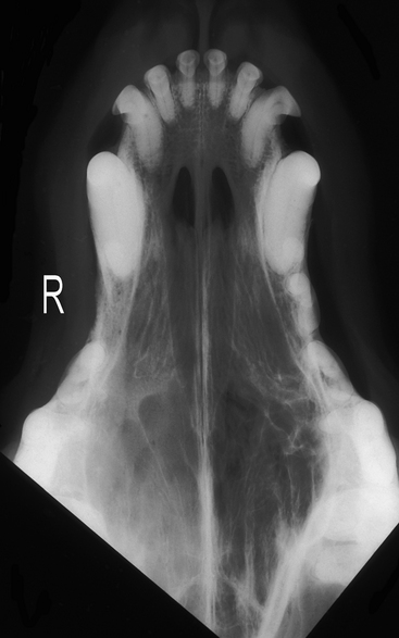
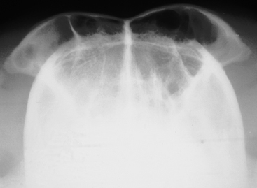
The most diagnostic skull radiographs are the open-mouth (dorsoventral or ventrodorsal intraoral) and the frontal sinus (rostrocaudal or “skyline”) views (Figs. 15-1 and 15-2). Left and right lateral oblique views are also typically evaluated. Radiographs of diagnostic quality require general anesthesia to ensure immobility.

Figure 15-1 Open-mouth ventrodorsal radiograph of a dog with an adenocarcinoma in the nasal cavity on the right side. Increased opacity of soft tissues may be seen in the caudal area of the right nasal cavity.
(Radiograph courtesy of the University of Florida.)

Figure 15-2 Frontal sinus skyline view of a dog with an adenocarcinoma of the right nasal cavity. Increased opacity of the right frontal sinus is evident.
(Radiograph courtesy of the University of Florida.)
Unfortunately, tumor-related radiographic abnormalities are not always different from those caused by infectious/inflammatory disorders. Common findings include destruction of bone, increased nasal and frontal sinus densities, often consistent with a mass lesion, and septum deviation/destruction. A nasal neoplasm is more often associated with soft tissue opacities and loss of turbinate detail, usually predominantly in one cavity; deviation of the nasal septum; evidence of bone invasion (lysis); and soft tissue/fluid opacities within the frontal sinus on the affected side. In contrast, an inflammatory or infectious rhinitis is more likely to be associated with localized soft tissue opacities and increased foci of lucency. Frontal sinus involvement is not typical. Generally, fungal rhinitis is characterized by intense turbinate lysis (lucency) with minimal increased soft tissue opacity.
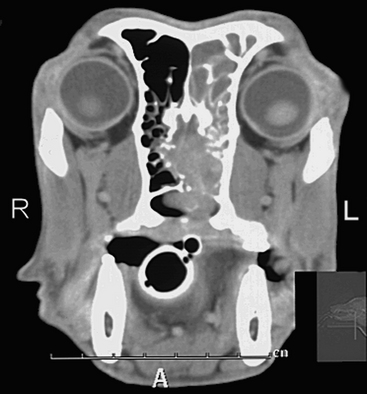
Plain radiography is an adequate screening test to rule out some causes of epistaxis or nasal discharge; however, advanced imaging is needed to assess the disease more completely. CT with and without contrast/enhancement is more accurate for determining the extent of local tumor infiltration than is plain radiography (Fig. 15-3). Furthermore, the length of survival is related to the extent of tumor involvement based on CT findings for dogs treated with radiation therapy.

Figure 15-3 Contrast-enhanced transverse computed tomography scan at the level of the caudal nasal cavity and rostral calvarium of a dog with a nasal adenocarcinoma. A large mass, visible in the left nasal cavity, has destroyed the turbinates and invaded the olfactory lobe and nasopharynx. The extent of tumor invasion into the brain would not be visible on radiographs.
(CT image courtesy of Dr. E. Riedesel, Iowa State University.)
Dogs with neurologic abnormalities referable to the central nervous system should undergo CT and/or MRI to determine the extent of invasion into the calvarium. Unfortunately, as with plain radiography, there is no pattern of CT abnormalities consistently associated with nasal and paranasal sinus neoplasms. CT guidance of the biopsy instrument enhances tissue sampling accuracy. In general, soft tissue details are better imaged with MRI than with CT. MRI is superior to CT and plain radiography for demonstrating the extent of tumor invasion into the frontal and olfactory lobes. Because CT or MRI provides a better view of tumors than plain radiography, it is a prerequisite for radiation therapy. Computed tomography findings are used to calculate the radiation dose and the field for radiation treatments in most treatment centers. CT findings can help in estimating the extent of radiation damage to normal tissues, particularly the eye.
